
بیماریهای قلب و عروق از شایعترین و در عین حال مهمترین مسائل حوزه سلامت هستند که آگاهی صحیح درباره آنها نقش بسزایی در پیشگیری، تشخیص زودهنگام و درمان مؤثر دارد. Many patients and even healthy people face daily common questions from cardiologists about heart health, warning signs, diagnostic methods and new cardiovascular treatments; But the correct answer to these questions requires expert opinion based on modern medical science.
In this regard, Dr. Majid Faraji, cardiologist and general medicine graduate from Iran University - cardiology graduate from Shiraz University of Medical Sciences - international cardiology fellowship (interventional cardiovascular procedures) from Tehran University - faculty member of Tehran University and responsible The angiography department of Gandhi Hospital, by participating in the "Doctor" television program, investigated and answered some of the common and frequent questions of heart and blood vessels. The content presented in this program is the result of clinical experience and specialized knowledge in the field of diagnosis, prevention and treatment of heart diseases and can be a reliable guide for the general public and heart patients.
In the rest of this article, the most important questions presented in the doctor program in the form of questions and answers about heart pain along with scientific and practical answers by Dr. Majid Faraji so that you can make more informed decisions with a clearer view of your heart health.
Check Some of the common questions of cardiology with the presence of Dr. Majid Faraji in the doctor program
In this section, we will examine some common and frequently asked questions in the field of cardiology, which were raised by the viewers of the doctor program. In this program, Dr. Majid Faraji, a cardiologist, answered these questions in a simple and scientific manner and provided important points about the diagnosis, prevention and treatment of heart diseases. In the following, you can read the explanation of these questions and specialized answers.
This case is one of the complications of angiography that occurs due to the injection of contrast material. During angiography, a contrast material is injected and a radiograph is taken at the same time. The contrast material causes an allergic reaction in some people. In a large percentage of patients, this problem is solved at the same moment in bed, but in a few people, delayed hypersensitivity reactions may also be seen. In this case, the swelling of the person's face is probably due to this reason, which is not worrying at all. This complication is usually solved with corticosteroid injections and anti-allergic drugs.
Usually, during the first and second injections, these sensitivities appear at the same time as the angiography, at the same moment these anti-allergic drugs are injected and the symptoms improve. For more details, you can read the article on See Before Angiography.
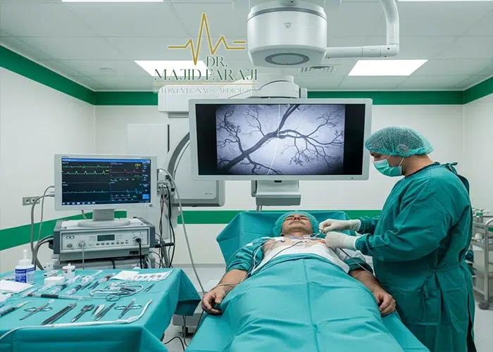
Actually, "angio" means "vessel" and "graph" means image and photo (or film). In general, the term angiography means seeing a vessel anywhere in the body. There are types of angiography of hand, foot, brain, kidney vessels, etc., but since Cardiac Angiography is a bit more common, more vital and sensitive, more heard about. In the event that every vein of the body can be angiographed.
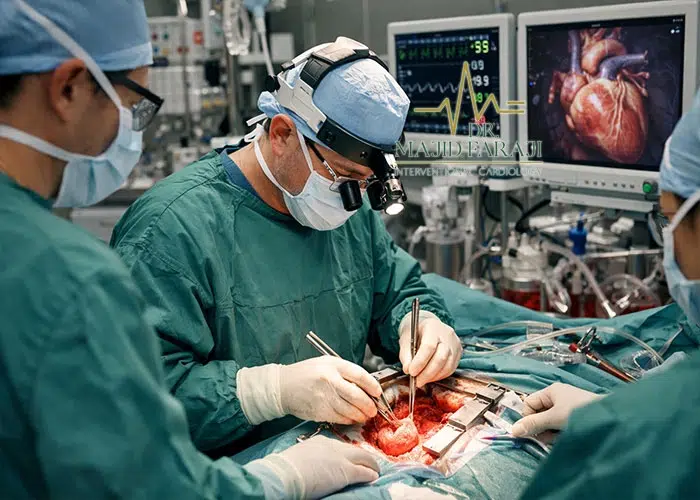
In fact, angiography There are two types of heart. One is an emergency procedure that is performed when a stroke occurs and we have to open the closed heart vessel in a short period of time. When a stroke occurs, time is of the essence. When a vessel is closed, all the cells supplied and nourished by that vessel are so-called suffocated. If this vein is opened in a suitable period of time, the cells will remain and the feeling of pressure and heaviness for the patient will be removed. This is because the blood supply to the cells is done properly. If this process happens with a delay, the cells die and the function of the heart decreases, which makes it difficult for the heart to pump.
For example, if angiography is performed after 24 hours, all the cells related to the vessel may be dead and the heart function will decrease. In other words, if the heart function or EF (Ejection Fraction) is 60% in a normal person, it becomes 20% in this person, which results in heart failure. This complication will cause problems such as shortness of breath during physical activity. In the heart and brain, after the cells die, complications occur that are irreversible.
The next group of patients who are candidates for angiography are patients who have not had a stroke, but have narrowed heart vessels. Symptoms begin when more than 70% of the vessel becomes blocked and narrowed. In this case, the person cannot easily do physical activity, fast walking, climbing the stairs, etc. In the case of these patients, a scan and CT angio (CTA) are performed, and after the narrowing of the arteries is determined, the person is a candidate for angiography.
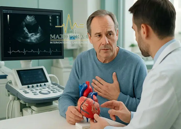
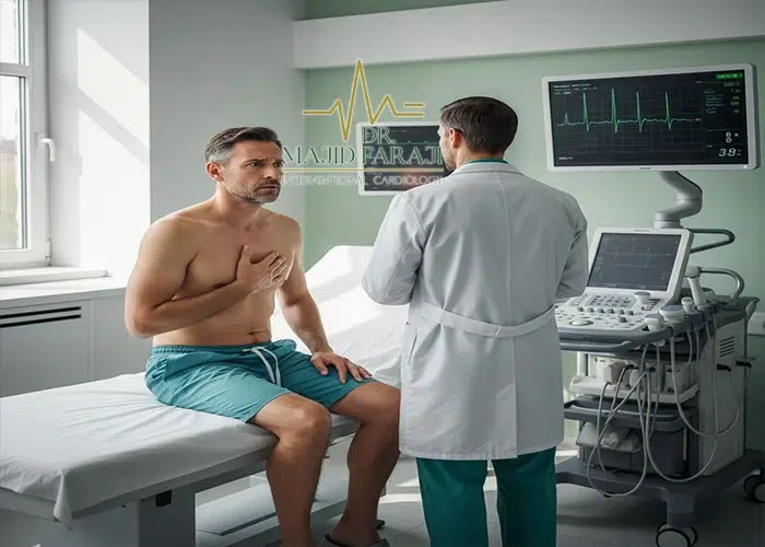
The reason for the heaviness and pressure of the chest must be the blockage of the heart vessel?
Not necessarily, for example, chest tightness, pain and pressure may also occur in people who are stressed or panicked. These states usually start while resting or when these people's thoughts and worries come to their mind. But heart problems usually cause symptoms with activity and doing something more than usual, such as climbing stairs or exercising, where there are usually no symptoms at rest, but with activity, symptoms appear. Therefore, pain and shortness of breath during exercise can be a sign of a heart problem.
Is Does youth and physical condition affect heart pain and pressure?
When a young person has low exercise capacity and has not exercised for a while, he may feel pain after exercise, but usually after exercising for a while, he will feel better and the body will return to normal. But someone who has a heart problem, even with the passage of time, still experiences symptoms.
Next How many years after the angiography, why does the doctor order to stop Plavix tablets?
The spring or stand that is placed in the vessel during angiography is made of metal and the body recognizes it as a foreign object. Initially, without medication, the blood can form a clot on the spring, so we dilute the blood with a combination of aspirin and Plavix to prevent clots from forming on the spring. After about a year, the body forms a layer on the spring that prevents blood from coming into direct contact with the spring. Therefore, at this stage, Plavix can be stopped and only aspirin can be continued.
Until a few years ago, angiography was usually performed through a leg vein, and problems such as hematoma (blood accumulation at the needle entry site) or movement restriction may occur. Today, most angiograms are performed through a vein in the hand. However, in some cases it is not possible to do it by hand, for example, when the vein in the patient's hand is tortuous or in women, when a heavy device enters the heart, the vessel may spasm and the passage of the device is closed.
Finally, whether from the hand or the foot, the path of access to the heart vein ends. One of the advantages of performing an angiography of the hand is that due to the limited space of the vessel, the bleeding stops faster and the puncture site is closed more easily. In the lower method, the bleeding may penetrate to the abdomen or the space around the leg and cause more problems.
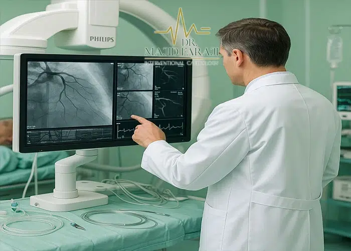
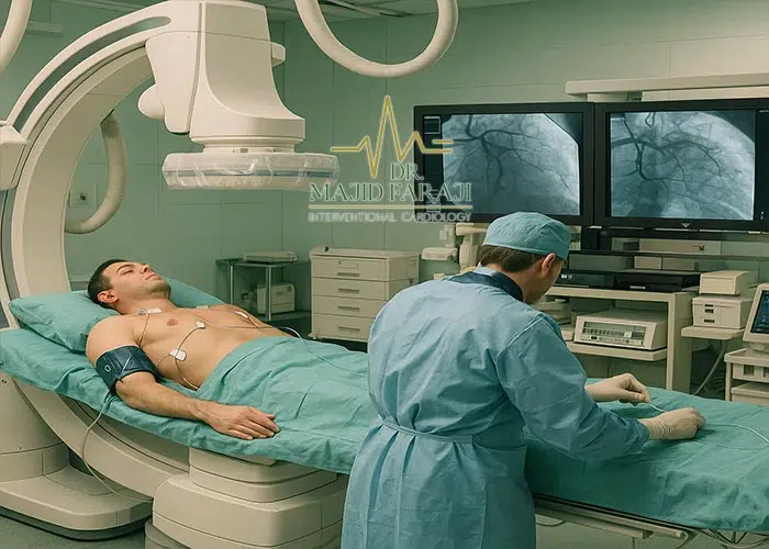
First, the doctor creates a way to enter the vein through the vein of the arm or leg. After that, a very thin and wire tube called a catheter is inserted through the same path and directed to the heart vessels. If the contrast agent is to be injected or a stent is placed, all these things are done through this catheter. This process is done under the X-ray machine and the doctor wears a lead suit. Contrast material is injected and images of heart vessels are recorded using a pedal. Because the arteries of the heart are three-dimensional and the images are two-dimensional, pictures are taken from different angles to get an accurate map of the arteries of the heart. Finally, these images are given to the patient on a CD.
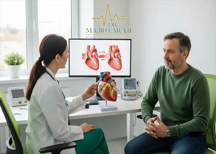
someone who performs angiography, what answers does he get from the doctor?
After the angiography, the doctor usually explains four situations to the patient:
- The vessels of the heart are healthy: In this case, the doctor says that the heart is healthy and no special treatment is needed.
- The arteries of the heart are narrow but do not need a spring: some narrowings are small and can be controlled with medicine. The reason these veins don't open is because the tiny heart veins usually supply less than 10 to 15 percent of the heart's blood supply and are no larger than 2 millimeters in diameter. Opening these veins usually does not have a significant effect and drug treatment is sufficient.
- an important and vital vein is narrowed: in this case, the doctor places a spring (stent) to open the vein and improve blood flow to the heart.
Cause What is leg pain after one year of angiography?
Leg pain after angiography is one of the side effects of this method. In some people, the nerve is affected, which causes pain, or a small clot forms, which causes long-term pain in the leg, which is a rare complication.
Is there an embolism during or after angiography?
No one can be sure that the risk is zero, because the physiology of the human body is different, just like when some people show sensitivity to contrast material.
Probability How much is death in angiography?
It cannot be said that it is zero, because this procedure is performed on the heart. The probability of death is about 1%, but in work experience, it usually occurs less than this number.
No, only local anesthesia with a low-volume insulin syringe in the arm or leg (entry site). No general anesthesia or other special anesthesia is necessary, because the veins are numb, and even if a few stents are placed in the patient's veins, it is usually not noticed.
The stent or spring is mounted on an inflatable balloon and prepared assembled. This spring has a specific length and diameter, which we determine based on angiography, what size the patient needs. Then we guide the stent mounted on the balloon into the vein and to the stenosis. When we reach the desired point, with a special device, we inflate the balloon with the appropriate pressure, for example, about 12 atmospheres. As the balloon inflates, the stent opens and sticks to the vessel wall. After that, we deflate the balloon and take it out, but the stent stays in place and keeps the vessel open.
next What happens to it in the body after the stent is installed?
When the stent is opened in the vessel and sticks to the wall of the vessel, it does not move anymore and remains fixed there. Over time, the inner tissue of the vessel grows over it and the stent practically becomes a part of the vessel. Even if a lot of pressure is applied to the body, the stent does not move. Also, if you pass through x-ray machines or inspection gates, it usually does not make a sound, because the volume of stent metal is very small.
The source of this article and the explanations provided is the video of Apparat Tabib, which is also available in the article. For information about cost of angiography and related questions, refer to the relevant article.