

پیشرفتهای پزشکی در دهههای اخیر باعث شده تشخیص بیماریها بسیار دقیقتر و کمتهاجمیتر از گذشته انجام شود. One of the most important achievements is endosonography or EUS; A method that is a smart combination of endoscopy and ultrasound and provides the possibility of observing the deep layers of the digestive system and the organs around it.
Many diseases of the pancreas, esophagus, stomach, intestines and even lymph nodes are difficult to detect with conventional imaging methods. This is where endosonography enters the field and helps doctors identify very small or deep lesions with high accuracy.
In this article, we have tried to explain in simple language but based on scientific sources what endosonography is, how it is performed, what are its uses and why it is considered one of the main pillars of advanced endoscopy today.
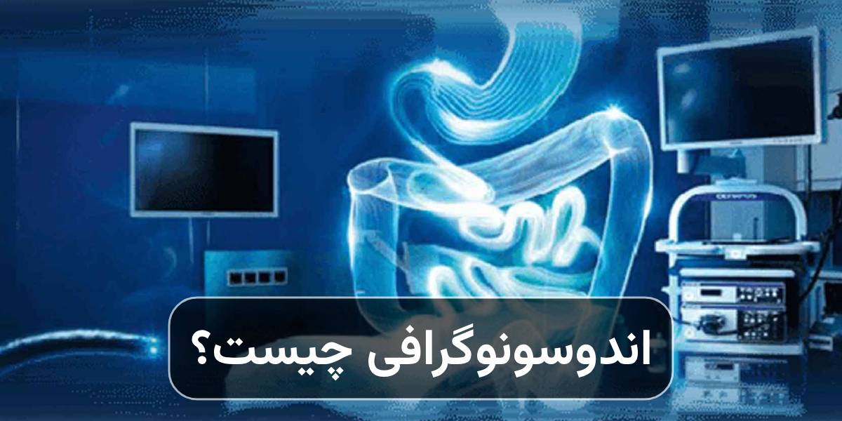
What is endosonography?
Endosonography (Endoscopic Ultrasound or EUS) is a diagnostic and therapeutic method in which a small ultrasound probe is placed at the tip of the endoscope. This device enters the body through the mouth or anus and simultaneously prepares endoscopic and ultrasound images of the tissues.
Unlike normal ultrasound that is performed on the skin, in EUS the source of sound waves is placed directly next to the desired organ; For this reason, the quality of the images is much higher and more detailed details of the wall of the digestive tract and the surrounding organs can be seen. Simply put, endosonography is a doctor's careful eye inside the body.
Why is endosonography important?
The importance of endosonography is that it can provide the following conditions:
- show the different layers of the wall of the esophagus, stomach and intestine separately
- Identify very small masses
- Determine the depth of waste penetration
- Check suspicious lymph nodes
- Possibility of accurate sampling (FNA or FNB)
According to the clinical reports published by the Mayo Clinic as well as the guidelines of the World Health Organization, EUS is considered one of the most accurate methods for evaluating gastrointestinal and pancreatic tumors.
How is endosonography performed?
The steps to perform endosonography are as follows:
- Patient preparation: The patient should usually fast for 6 to 8 hours. If the bowel is examined, bowel cleansing may also be necessary.
- Anesthesia or sedation: Most patients undergo intravenous sedation so that they do not feel discomfort during EUS.
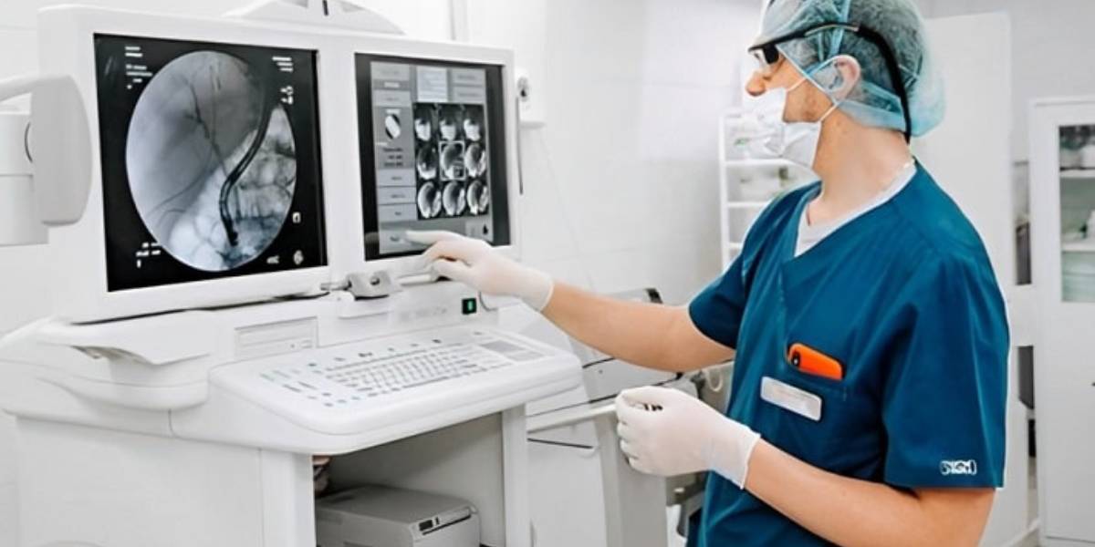
- Entering the device: an endoscope equipped with an ultrasound probe is inserted through the mouth (for the esophagus, stomach and pancreas) or through the anus (for the rectum and terminal colon).
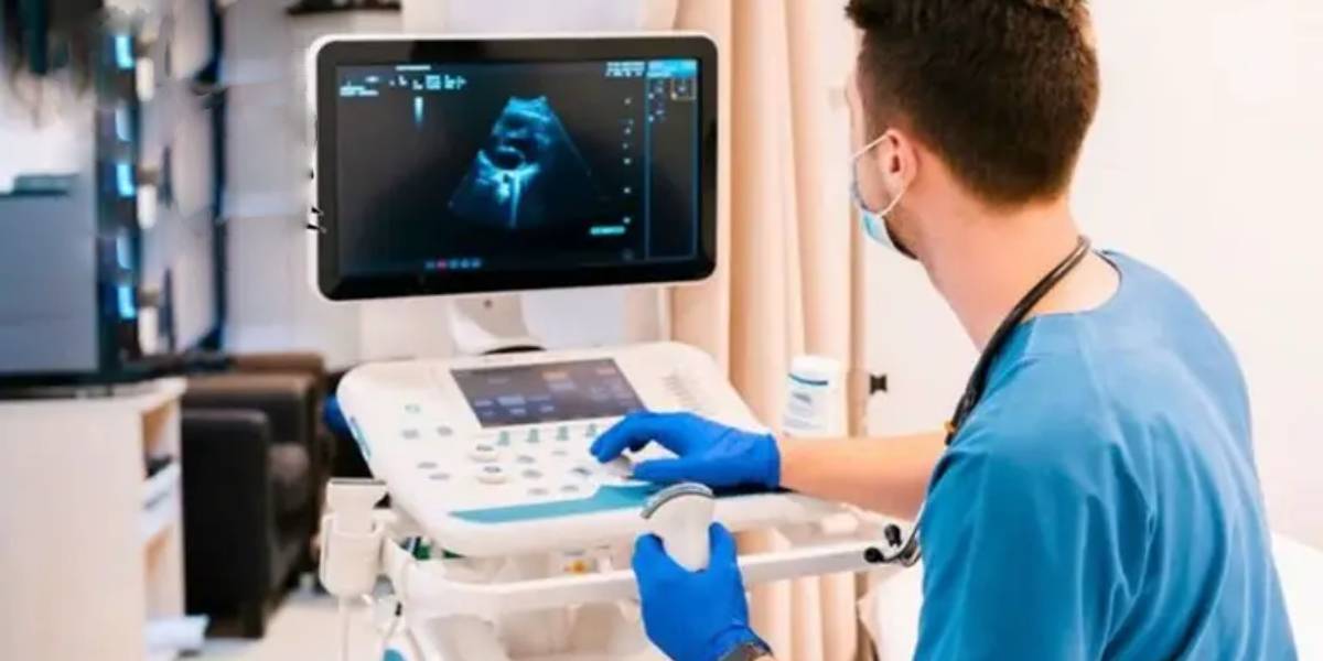
- Precise imaging: With the slow movement of the device, the doctor observes layer-by-layer images of the digestive wall and adjacent organs.
- Sampling if needed: if a suspicious lesion is seen, a sample can be taken from it with a special needle; No need for open surgery.
The main applications of endosonography
The main uses of endosonography are as follows:
Inspection of pancreatic diseases
EUS is the most accurate method for diagnosing the following:
- Pancreatic tumors Pancreatic cysts
- Chronic inflammation of the pancreas
Especially when the CT scan or MRI did not give definitive results.
Staging of gastrointestinal cancers
Endosonography determines the following:
- How deep has the tumor penetrated?
- Whether the lymph nodes are involved or not
This information is vital to choose the best treatment method.
Evaluation of submucosal masses
Some lumps are located under the surface of the mucosa and can only be seen as bumps with normal endoscopy. EUS determines the exact source of these masses (muscle, fat or nerve tissue).
Targeted sampling (EUS-guided biopsy)
One of the most important advantages of endosonography is the possibility of accurate sampling of deep lesions without surgical incision.
Therapeutic applications
Today, EUS is not only a diagnostic tool, but is also used in some therapeutic procedures, including the following:
- Evacuation of cysts
- Drainage of abscesses
- Injecting medicine into masses
- Helping to treat persistent pancreatic pain
Endosonography next to advanced endoscopy
Endosonography is part of a larger set of advanced endoscopy; A branch of medicine that, in addition to observation, also provides the possibility of treating lesions.
In this field, techniques such as EMR and ESD are used to remove superficial lesions of the digestive tract. Knowing The difference EMR and ESD is very important to choose the right method:
- EMR is usually used for smaller and more superficial lesions.
- ESD allows larger pieces of waste to be removed, but is a more complex technique.
Endosonography is often performed before these procedures to determine the depth of the lesion and make a more accurate treatment decision.
Advantages of endosonography compared to other imaging methods
The most important advantages of endosonography are as follows:
- Very high accuracy in detecting small lesions
- Direct observation of the layers of the digestive wall
- Possibility of simultaneous sampling
- No need for open surgery
- Less side effects compared to invasive methods
Is endosonography dangerous?
EUS is generally considered a safe procedure. However, as with any medical procedure, there is a possibility of rare side effects, including the following:
- Temporary sore throat
- Bloating or feeling full in the stomach
- Slight bleeding at the sampling site
- Rarely infection or perforation
The incidence of these complications is very low, especially when performed by an experienced doctor.
Who are suitable candidates for endosonography?
Endosonography is usually recommended for people with the following conditions:
- They have a suspicious lesion in endoscopy or CT scan
- The possibility of pancreatic tumor or complex cysts is considered
- They need accurate cancer staging
- They have submucosal masses
- The cause of chronic abdominal pain is unknown
Necessary preparations before doing EUS
The most important preparations before doing endosonography are as follows:
- Fasting for at least 6 hours
- Informing the doctor about blood thinners
- Bringing previous imaging documents
- The presence of a companion to return home
Endosonography is one of the most accurate and advanced diagnostic tools in modern medicine, which enables in-depth examination of the digestive system and its surrounding organs. This method plays a key role in early detection of cancers, evaluation of suspicious masses and treatment planning, and today it is considered an integral part of advanced endoscopy.
Combining EUS with techniques such as EMR and ESD (with a proper understanding of the difference between EMR and ESD) allows physicians to treat many lesions without open surgery; An issue that has significantly improved the quality of life of patients. If the doctor has prescribed endosonography for you, there is no need to worry; This method is usually safe, accurate and very helpful in reaching the correct diagnosis.