
Heart scan with spect and perfusion method is one of the accurate and non-invasive methods of diagnosing heart diseases, which helps to show heart muscle and coronary artery problems in time. Due to the high prevalence of heart diseases and their role in global deaths, this type of cardiac imaging has become an essential tool in timely diagnosis and effective treatment planning. This test is performed under the supervision of a cardiologist or a nuclear medicine doctor and can provide important information about heart function and the risk of vascular occlusion.
Table of contents
- 1 What is cardiac axon by SPECT and perfusion method?
- 2 The best heart SPECT scan clinics in Tehran
- 2.1 ✅ Important note:
- 2.2 7 important stages of heart scanning with the SPECT and perfusion method
- 2.3 What is the importance of SPECT and perfusion scanning in the diagnosis of heart disease?
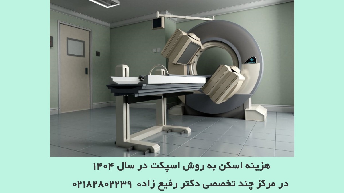
- 2.4 How much does SPECT scanning cost?
- 2.5 Specialists' answer to SPECT scan is it dangerous or not?
- 2.6 Who should not undergo SPECT heart scan?
- 2.7 ✅ Advantages of cardiac scanning with SPECT method compared to angiography
- 2.8 1. Non-invasiveness
- 2.9 2. Fewer risks
- 2.10 3. No need for hospitalization
- 2.11 4. Lower cost
- 2.12 5. Diagnosis of ischemia and heart muscle function
- 2.13 6. Suitable for high-risk patients
- 2.14 7. Easier preparation
- 3 ✅ Summary
What is heart axon by spect and perfusion method?
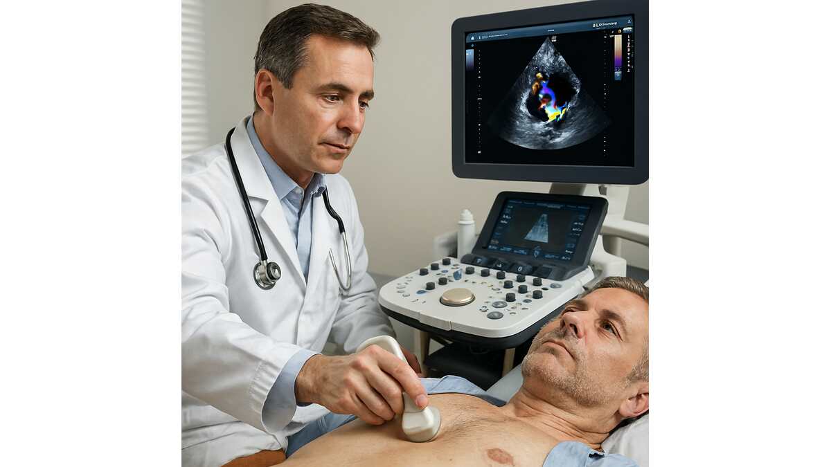
acardiac xin with the aspect method and perfusion is an imaging test that uses radioisotopes and the device SPECT examines the function of the heart and the blood supply to the heart muscle. This method helps doctors to identify areas of the heart that have less blood flow and to treat early if there is a blockage in the coronary arteries. This test is non-invasive and painless and is usually used in the accurate diagnosis of coronary artery diseases and heart function evaluation.

The best clinics for heart SPECT scan in Tehran
Below is a list of the best clinics and centers to perform heart scanning with the Spect and Perfusion method in Tehran . These centers have advanced devices, experienced staff and clinical experience and provide cardiac imaging services with SPECT and myocardial perfusion well.
| Clinic name | Specialty | Geographical location | Contact number | Important features | |
|---|---|---|---|---|---|
| Vistan Clinic | SPECT heart scan, myocardial perfusion, imaging Nuclear | AriaAll nuclear medicine services, heart SPECT scan | Vank / Saadat Abad | – | SPECT device with CT Fusion capability Experienced staff Cooperation with major hospitals |
| Heart Clinic Iran | Non-invasive diagnosis of diseases Cardiac | Fershetha area | – | SPECT scan + stress test before and after consultation online registration | |
| Olympia diagnostic center | Diagnostic services including MRI, CT, SPECT and... | Felake Roberto area - Blvd. Keshavarz | – | Equipped laboratory Admission without insurance booklet Cardiac tests during the day | |
| Dr. Alizadeh Clinic - Nuclear Medicine | Nuclear imaging of the heart and body | Vali Asr region | – | Expert based in Location Personalized service Flexible scheduling |
Important note:
- Vistan Clinic as one of the leading centers in the field of heart scanning with the Spect and Perfusion method, due to its advanced devices, calm and suitable environment and person-oriented services, is a very suitable option for people who are looking for high accuracy and convenience in performing the test.
7 important stages of heart scanning with the spect and perfusion method
7 important steps for heart scanning with SPECT and perfusion method that you should know are:
- Patient preparation
One of the most important steps in successfully performing a heart scan with the SPECT and perfusion method is the correct preparation of the patient. Preparation includes several important parts:
- Following a special diet: The doctor may ask you to avoid eating and drinking for a few hours before the scan (usually 4 to 6 hours).
- Not consuming caffeine: Caffeinated drinks such as tea, coffee, soft drinks and even chocolate may interfere with the scan.
- Discontinuation of certain medications: Some cardiac medications should be temporarily discontinued. Of course, any change should be made only according to the doctor's opinion.
- Mental and mental preparation: Many patients have concerns about this scan, but you should know that this procedure is safe, painless and non-invasive.
At this stage, a harmless radioactive substance such as technetium or thallium is injected into the patient's vein. This substance enters the heart through the blood flow and accumulates in the parts of the heart muscle that have good blood flow. This substance acts like a marker and helps the machine to evaluate the heart's function more accurately.
Radioactive materials used in this method have a very small dose and are safe for the body. Even in many cases, patients can return to their daily activities after doing a heart scan with the Spect and Perfusion method.
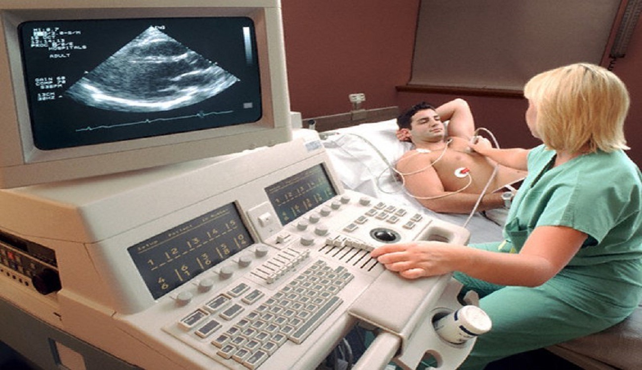
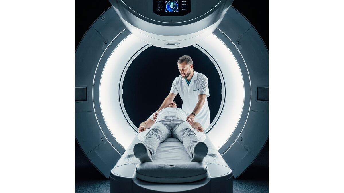
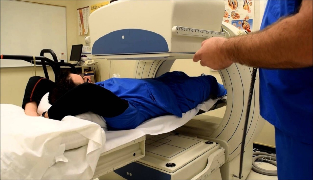
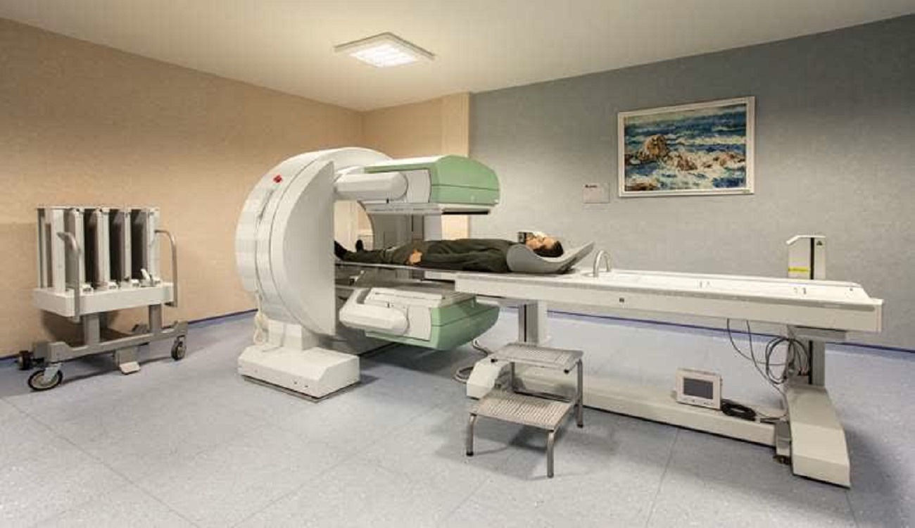
3. Imaging at restAfter injecting the radioactive substance, the patient is transferred to the imaging room. At this stage, the patient lies comfortably on the bed of the Spect machine. The device rotates around the patient's body and transmits the information to the image processing system.
These images show how blood flows through the heart muscles when the heart is at rest. The presence of points with less blood supply can be a sign of damage or heart disease.
4. Creating conditions of stress or activityTo check the function of the heart in conditions of activity or stress, the heart should be put in an overworked state. This is done in two ways:
- Physical method: Walking on a treadmill or stationary bike under the supervision of a doctor. This method is usually suitable for people who are capable of physical activity.
- Drug method: In patients who cannot exercise, drugs are injected that have the same effect as exercise on the heart, such as increasing heart rate and blood pressure.
The purpose of this stage is to check the heart's performance when the need for oxygen and blood increases. Usually, the radioactive material is also injected in this second stage so that imaging can be done after stress.
5. imaging after stressAfter the end of the stress phase, the patient is prepared for imaging again. This stage is similar to the imaging stage at rest, with the difference that now the heart is active and needs more blood.
Comparing the images of the rest and stress phase can show the doctor whether the blood supply has changed in different conditions. For example:
- Areas that have blood supply at rest, but not during stress, may have blood clots.
- Areas that do not have blood supply in any of the stages, are probably permanently damaged.
The recorded images should be interpreted by a specialist or cardiologist. Using these images, the doctor can:
- Determine the extent of heart muscle damage.
- Check the condition of heart pumping function.
- Determine the severity or location of blood vessel blockage.
- decide whether the patient needs drug treatment, angiography or other procedures or not.
After interpreting the results, the doctor determines the appropriate treatment plan for the patient. At this stage, decisions such as the following may be made:
- Starting or changing cardiac medications
- Advice to change lifestyle (diet, smoking cessation, exercise)
- Refer to angiography or surgery
- Periodic checks with scans or other tests

Myocardial perfusion scans are a widely used procedure to diagnose ischemic heart disease. This activity reviews the importance of these scans and highlights the role of the interprofessional team in performing them.
- Identify the indications for a myocardial perfusion scan
- Assess the technique of performing a myocardial perfusion scan.
- Evaluate the clinical significance of myocardial perfusion scan.
- Communicate how an interprofessional team approach can improve patient outcomes when using myocardial perfusion scanning on appropriate patient populations.
Myocardial perfusion scan is a widely used method for diagnosing ischemic heart disease. This activity examines the importance of these scans and highlights the role of the interprofessional team in performing them.
Objectives: Identify the indications for a myocardial perfusion scan
Evaluate the technique of performing a myocardial perfusion scan.
Evaluate the clinical importance of a myocardial perfusion scan.
How to use a myocardial perfusion scan in the appropriate patient population, how an interdisciplinary team approach can improve patient outcomes.
Quoted from the site: www.ncbi.nlm.nih.gov
What is the importance of spect and perfusion scan in the diagnosis of heart disease?
Below is a table of the importance of scanning of the heart with SPECT and perfusion in the diagnosis of heart diseases:
| Title | Description |
|---|---|
| Method name | SPECT scan / myocardial perfusion |
| Main goal | Evaluation of blood flow to Heart muscle (myocardium) at rest and under stress |
| Diagnostic use | - Diagnosis of hyperemia (decreased blood flow) in patients with cardiac sector - Determining the size of the affected area in myocardial infarction - Prediction of cardiac risk (such as heart attack) - Assessment of heart function after stroke or treatment |
| Method Perform | – Radiopharmaceutical injection (such as Tc-99m MIBI or Tetrofosmin) – SPECT/CT imaging before and after stress (exercise or drug) – Comparison of rest and stress images to find areas of low blood flow |
| Advantages | – Non-invasive and relatively safe – Ability to accurately detect areas with reduced blood flow blood – help decide whether angioplasty or surgery is needed – follow up patients after treatment |
| disadvantages | – small amount of radioactive radiation – long time to perform the test (several hours) – results may be affected by factors such as obesity, diabetes, or cardiac dysfunction |
| sensitivity and Characteristic | – Sensitivity: 80–90% – Specificity: 70–85% in the diagnosis of coronary artery disease |
| Comparison with other methods | – vs coronary CT: higher cost and radioisotope radiation, but direct image of myocardial blood flow – vs stress MRI: lower cost and higher availability in many Centers |
| Sample results | – Ischemic area: decreased blood flow under stress that improves at rest – Necrotic area (replaced): decreased radioisotope uptake at both rest and stress |
Heart scan with spect and perfusion method Myocardium is one of the most accurate non-invasive methods for diagnosing heart diseases, especially coronary artery disease, which is able to identify the blockage, its location, severity and type.

What is the cost of scanning with the Spect method?
The cost of heart scanning with the aspect and perfusion method depends on various factors such as the type of device, the location, the radioisotope materials used and It depends on the clinical expertise of the examination center. In our country, the cost of this test usually ranges from 1 to 3 million tomans. Of course, it should be noted that performing this test in well-equipped centers with experienced staff greatly increases the accuracy of the results.
Vistan Clinic is one of the reliable and experienced centers in the field of performing heart scanning with the Spect and Perfusion method. With the latest generation of cardiac imaging devices, medical staff and expert technicians, and a hygienic and soothing environment, Vistan Clinic is one of the trusted destinations for accurate diagnosis of heart diseases and heart scanning with Spect and Perfusion methods without the need for invasive methods.
This clinic is located in Tehran city and provides diagnostic services of heart scanning with the spect and perfusion method in full compliance with international standards and with a person-oriented approach. At Vistan Clinic, people can perform a heart scan with the Spect and Perfusion method according to the insurance book and only with the referral of the attending physician.
To sum up, spect and perfusion heart scanningbesides high accuracy in the diagnosis of heart diseases, it also provides the possibility of proper planning for treatment, and choosing Vistan Clinic as a reference center in this field can guarantee the quality and accuracy of the results.
Marzdaran branch address:
Tehran - Ashrafi Isfahani highway - on Marzdaran boulevard - after Rezainejad street - corner of Bahar street - No. 184 - Iman building - east ground floor (behind Dr. Khatami's pharmacy)
Saadatabad branch address:
Tehran - Saadatabad - north-west side of Sarro intersection (intersection of Paknejad and Saro) - beginning of Nami street - No. 19 - next to +10 police station - top floor of Dr. Dashblaghi pharmacy
To find out about appointment conditions and coordination for spect and perfusion heart scan, you can call the clinic phone number 09966333097 to get the necessary guidance.
Experts' answer to Scan Aspect, is it dangerous or not?
According to cardiology and nuclear medicine experts, cardiac SPECT scanning is generally considered a safe and procedure, especially when performed by experienced personnel and in well-equipped centers. Of course, as with any medical experiment, there may be mild side effects or small risks, which we will discuss below.
Dangers and possible complications of heart SPECT scan:
- Receiving a small amount of radiation:
- In this method, radioactive substances (such as technetium-99m) are used, whose dose is very low and is equivalent or even lower than other imaging methods such as CT scan.
- Radiation risk in this case is very small and normal and does not cause problems for most patients.
- Complications caused by stress test (if performed):
- If the scan is accompanied by a pharmacological stress test (such as dopamine, dipyridamole or adenosine), the person may have symptoms such as:
- Headache
- Heartbeat
- shortness of breath
- feeling of heat or burning in the body
- Experience low blood pressure, which is usually short-lived and without serious consequences.
- If the scan is accompanied by a pharmacological stress test (such as dopamine, dipyridamole or adenosine), the person may have symptoms such as:
- Rare allergic reaction:
- Allergic reactions to radioisotope materials or stress medications are very rare, but if there is a history of allergies, the doctor should be informed before the test.
- Danger for pregnant and lactating women:
- This test is not recommended during pregnancy, because radiation may be transferred to the fetus.
- Breastfeeding mothers may also need to temporarily stop breastfeeding. In these cases, consultation with a doctor is necessary.
Who should not have a cardiac SPECT scan?
- Pregnant
- People with severe sensitivity to stress medications (with prior notification)
- Persons with severe heart or respiratory failure(if not approved by the doctor)
Important note: If you intend to perform a heart scan with the Spect and Perfusion method, inform the treatment staff of all your medical history, pregnancy, breastfeeding and drug allergies before the test.
Advantages of cardiac scanning with SPECT method compared to angiography
✅ Advantages of cardiac scanning with SPECT method compared to angiography
Heart scan with SPECT (Single Photon Emission Computed Tomography) and coronary angiography are both used to diagnose coronary artery diseases, but they have important differences in terms of type of method, risks, accuracy, cost and how to do it. Below we discuss the advantages of SPECT scanning over angiography:
1. Being non-aggressive
- ✅ SPECT: It is completely non-invasive and only involves a simple injection and imaging.
- ❌ angiography: It is an invasive procedure in which a catheter is inserted into the body through the shoulder or thigh artery and reaches the heart.
2. Less risks
- ✅ SPECT: It has very low risks, only mild reactions such as headache or heart palpitations may occur in the stress test.
- ❌ angiography: has more risks such as:
- Bleeding at the catheter site
- Allergic reaction to contrast agent
- cardiac arrhythmia
- Heart or stroke (in rare cases)
3. No need for hospitalization
- ✅ SPECT: It can be done without hospitalization and even in one day.
- ❌ angiography: requires post-operative care and usually a few hours of hospitalization.
4. Lower cost
-
✅ SPECT: It costs less than angiography.
- ❌ angiography: due to the need for operating room, catheter, anesthesia and expert staff, it is expensive.
5. Diagnosis of ischemia and heart muscle function
- ✅ SPECT: It is able to show areas with low blood flow (ischemia), dead areas (necrosis) and residual heart activity.
- ❌ angiography: it only shows blockage of vessels and does not give information about the functioning of the heart muscle.
6. Suitable for high-risk patients
- ✅ SPECT: For patients who cannot exercise on a treadmill, it can be done with stress medication.
- ❌ angiography: it is more dangerous for patients with severe history of heart disease.
7. Easier preparation
- ✅ SPECT: only a few hours of fasting and cutting caffeine are needed.
- ❌ angiography: more detailed preparation and more care before and after the test is required.
Summary
| Criteria | SPECT scan | Angiography |
|---|---|---|
| Type of method | Non-invasive | Invasive |
| Risks | Many Lower | Higher |
| Need hospitalization | No | Yes |
| Cost | Lower | Higher |
| Information about muscle function Heart | Yes | No |
| Suitable for high-risk patients | Yes | No |
| Diagnosis of vascular occlusion | Yes (with good accuracy) | Yes (more accurate) |
Important note:
- SPECT scan is very suitable for screening, risk assessment, ischemia area determination and treatment planning .
- Angiography is a better choice when direct imaging of the coronary arteries is needed and angioplasty or bypass treatment is being considered.
If you are looking to decide which method to choose, consultation with a cardiologist and nuclear physician is essential. Vistan Medical Group are the best specialists and consultants to perform heart scan with Spect and Perfusion method.

Frequently asked questions about cardiac scanning with SPECT and perfusion method
What is a SPECT scan of the heart?
It is a non-invasive imaging method to check the blood flow of the heart muscle (perfusion) and diagnose coronary artery blockage.
Is this experiment dangerous?
It has very little risk and is generally safe; Mild side effects such as headache or heart palpitations may occur.
How long does it take?
About 4 to 6 hours, including two stages: at rest and under stress
Who should do this test?
People suspected of having coronary artery disease, cardiac ischemia, or those who need to evaluate heart function after a stroke.
When will the result be ready?
Usually 1-2 working days after testing.
Is stress medication necessary?
Sometimes yes, especially if the person cannot run on a treadmill, stress medications are used instead of exercise
Will I get radiation?
Yes, you receive a very small amount of radiation that is harmless to the body.
What should be done before the test?
Do not eat/drink anything 4 to 6 hours before the test, tea, coffee and smoking are also prohibited.
Conclusion
Heart scan with spect and perfusion method is one of the most accurate and widely used non-invasive imaging methods for diagnosing heart diseases. This test helps doctors not only identify the presence of blockage in the coronary arteries, but also determine its location, severity, and type. In this article, we tried to introduce you to the heart scan using the spect and perfusion method and pay attention to the important points and methods of performing this scan.