
In the advanced era of cardiac imaging, echocardiography and Doppler TDL Fellowship Echo are prominent as one of the pillars of diagnosis, monitoring and guidance of cardiac treatment. These techniques, by combining ultrasound image, Doppler probe, tissue imaging (Tissue Doppler / TDL) and complex analysis, allow accurate evaluation of the structure, function and hemodynamics of the heart. Specialized learning in this field, especially at the level of super-specialist fellowship, requires mastery of physical, technical, clinical and managerial concepts. In this article, six special points about Echocardiography and TDL Doppler Echo Fellowship are discussed, which are essential for Echo Fellowship specialists and interns.
Table of contents
- 1 Specialized echocardiography - physics, technique and application
- 2 Brief comparison of specialized echocardiography techniques
- 2.1 Preoperative echocardiography - role in preoperative assessment
- 2.2 Specialist points in evaluation Preoperative
- 2.3 Where is the best echocardiography clinic in Tehran?
- 2.4 Some echocardiography centers in Tehran
- 2.5 Key indicators of preoperative echocardiography
- 2.6 Tissue Doppler echocardiography (Tissue Doppler) – myocardial motion analysis
- 2.7 The concept of Doppler tissue echocardiography
- 2.8 Color echocardiography - color Doppler and hemodynamic applications
- 2.9 TDL echocardiography (Fellowship) – Subspecialty stage
- 2.10 Subspecialty echocardiography – future and new trends
- 2.11 Echocardiography and Doppler TDL Fellowship Echo What is it?
- 2.12 What is the purpose of echocardiography and Doppler TDL?
- 2.13 What is the difference between normal echo and TDL echo?
- 2.14 Is TDL echo painful?
- 2.15 What is the difference between TDL and color Doppler?
- 2.16 When does a doctor recommend a TDL echo?
- 2.17 How are TDL echo results interpreted?
- 2.18 Fellowship What is echo?
- 2.19 Can TDL echo be done for all patients?
- 2.20 What is the future of TDL echocardiography?
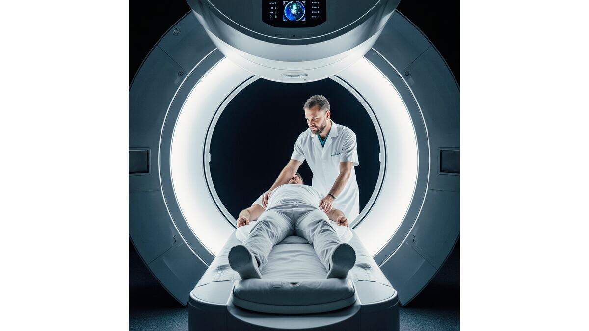
Specialized echocardiography - physics, technique and application
Definition and importance
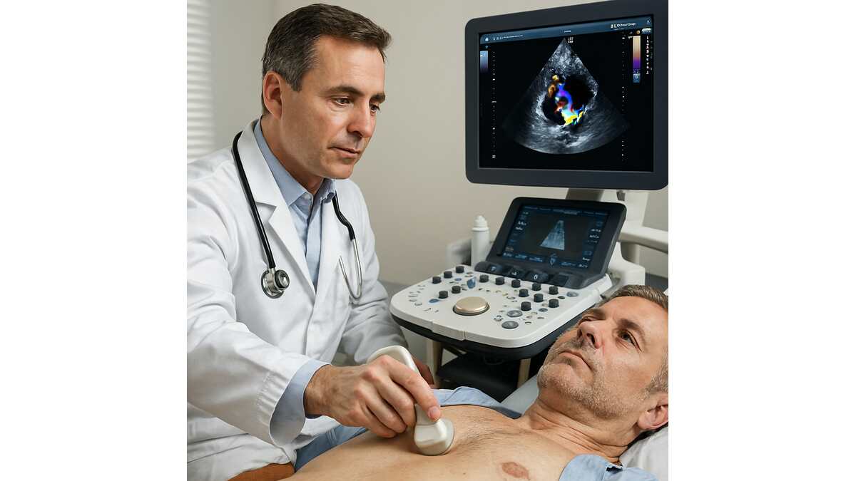
At the specialized level, Echocardiography and Doppler Echo Fellowship TDL means using more advanced techniques Echocardiography – includes 2D, 3D imaging, color Doppler, pulse-wave Doppler, tissue Doppler (TDI/TDL) – for clinical and research evaluation of the heart.
The inclusion of these techniques in the Echo Fellowship means the ability to provide accurate reports, early diagnosis of disorders cardiac function is to guide treatment and monitoring after interventions.
Physical and technical components
- Principles of ultrasound and sound waves: familiarity with probe frequency, sound absorption, reflection coefficient, radiation angle, temporal and spatial resolution.
- Doppler of blood flow and tissue: in classic Doppler, the speed of blood flow and its direction are checked; In tissue (TDI/TDL) myocardial tissue velocity is evaluated. Device settings: probe selection, radiation angle parallel to the flow as much as possible, scale, gain, removal of artifacts, selection of standard and non-standard views.
- Standardization of the report: familiarity with quantitative techniques such as measuring wall thickness, cavity volume, Doppler tissue speed (S', E', A'), ratios and performance indicators.
Specialized Applications
Echocardiography and Doppler TDL Fellowship Echo In the field of specialized echocardiography, applications such as evaluation of complex valvular diseases, cardiomyopathies, pulmonary arterial hypertension, preoperative and postoperative stages, and echo-guided methods are proposed. Therefore, the fellowship specialist must be able to accurately diagnose and suggest management.

Overview comparison of specialized echocardiography techniques
In the table below, a summary of specialized echocardiography techniques has been compiled regarding Echocardiography and Doppler TDL Fellowship Echo.
| Technique | Main index | Key application |
|---|---|---|
| Two-dimensional (2D) | Volume, thickness, wall movement | Basic function of the heart, Anatomy |
| Three-Dimensional (3D) | More detailed volume, complex geometry | Valves, right ventricle, research studies |
| Color Doppler / blood-acceleration | Blood flow direction and velocity | Valvular insufficiency, Shunts |
| Pulse-wave Doppler (PW) | Velocity at specific point | Filling pressure, valve flow |
| Tissue Doppler (TDI/TDL) | Myocardial velocity | Dystolic dysfunction, prediction |
Preoperative echocardiography – role in preoperative assessment
Importance and role
In planning cardiac surgery or non-surgical interventions, Echocardiography and Doppler TDL Fellowship Echo play a vital role. From identifying anatomy, estimating risk, evaluating left and right ventricular function, to evaluating preoperative hemodynamic conditions, all can be done through these techniques.
Expert tips in preoperative assessment
- Examination of left ventricular function (LVEF), wall thickness, wall movement, diastolic indices such as E/A, E/E' using Doppler tissue.
- Evaluation of valves (aortic, mitral, tricuspid, pulmonary): stenosis, regurgitation, pressure and flow parameters, using color Doppler and pulse-view.
- Identify right ventricle, pulmonary artery pressure, and associated problems such as pulmonary fibrosis or pulmonary embolism that can increase the risk of surgery.
- Determining the amount of risk based on echo and Doppler findings: for example, poor right ventricular function or high pulmonary artery pressure may require special preoperative interventions.
- Communication with the anesthesia team and the surgeon: the echo report should include a comprehensive picture and recommendations for preoperative management, discharge and postoperative monitoring.
Where is the best echocardiography clinic in Tehran?
Vistan Clinic is one of the advanced multi-specialty centers for Echocardiography and tdl Doppler Fellowship Echo in Tehran, with the use of expert medical staff and Modern diagnostic and treatment equipment provides various services in medical fields. In this center, there are departments such as cardiology, gastroenterology, neurology, physical medicine and rehabilitation, diagnostic imaging, etc.
Among the strengths of Vistan Clinic, we can mention proper location in the west of Tehran (Marzdaran and Saadat Abad branches) and easy access.
Advantages and notable points Echocardiography and Doppler TDL Fellowship Echo at Vistan Clinic:
- Using up-to-date equipment for diagnosis and treatment;
- Experienced and multispecialty medical team;
- The possibility of receiving various services in one center (saving time and energy of the client);
- Modern and user-friendly environment.
Tips for clients: - When visiting, be sure to make an appointment so that you are less delayed;
- If you intend to have specialized heart or echocardiography services, contact the clinic in advance to coordinate the relevant department;
- Bring previous medical documents (if you have them);
- It is better to ask if the center cooperates with certain insurances or not (if you have insurance).
Vistan clinic address: Tehran, Sadat Abad, northwest side of Saro intersection (intersection of Paknejad and Saro)-next to Dasht Behesht complex
Vistan clinic contact numbers for making appointments and reserving appointments in the field of Echocardiography and Doppler TDL Fellowship Echo: 02191090775 – 02128423424 – whatsapp

some echocardiography centers in Tehran
In the following, I have prepared a brief table of some of the best echocardiography and Doppler TDL fellowship echo centers in Tehran
| Name of the center | Type (clinic / hospital) | Short description | Contact number |
|---|---|---|---|
| Vistan clinic | Specialized clinic | Has specialized echocardiography departments | 09966333097 – 09339198680 |
| Tehran Heart Center Hospital (Tehran University of Medical Sciences) | Specialized hospital | Having an echocardiography department specialized in heart and blood vessels. | – |
| Tehran Arrhythmia Clinic (Tovanir Square) | Specialized heart clinic | Providing echocardiography services, including specialized heart echoes. | – |
| Shahid Rajaee Heart Hospital, Tehran | Specialized hospital | One of the reputable heart centers that is seen in the list of heart echo centers | – |
| Lovasani Hospital Tehran | specialized hospital | advanced heart center in Tehran which also includes echocardiography department. | – |
| specialized heart clinic of Dr. Najafi | private clinic | one of the specialized heart centers including echocardiography services diagnostic | – |
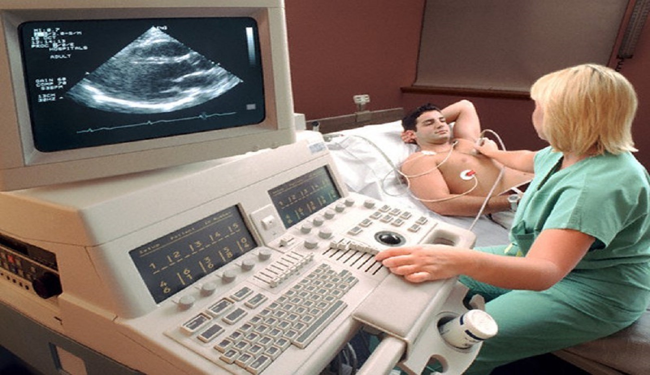
Echocardiography is a standard modality to diagnose DCM. It involves detecting structural and functional changes in the LV and excluding other CVD that could be the potential reason for those changes in a patient with DM. Comprehensive evaluation of DCM with standard transthoracic Doppler echocardiography involves the assessment of diastolic dysfunction, LV filling pressure and myocardial perfusion reserve.
Echocardiography is a standard method to diagnose DCM. This method includes the diagnosis of structural and functional changes in the left ventricle and the rejection of other cardiovascular diseases that can be the potential cause of these changes in a patient with diabetes. Comprehensive assessment of DCM with standard transthoracic Doppler echocardiography includes assessment of diastolic dysfunction, left ventricular filling pressure and myocardial perfusion reserve.Quoted from the site: sciencedirect.com
Key indicators of preoperative echocardiography
| Index | Alarm threshold | Interpretation |
|---|---|---|
| LVEF less than 30% | High risk | Special monitoring may be required Supportive interventions are necessary |
| E/E’ > 15 | High left ventricular filling pressure | Higher risk of postoperative complications |
| Pulmonary artery pressure (PASP) > 50 mmHg | Right ventricle at risk | Increased possibility of postoperative complications finds |
| severe aortic valve regurgitation + reduced LVEF | risky combination | requires further investigation and possibly concurrent interventions |
Fellowship Recommendations
- Preparation of preoperative echo protocol: including standard views, summary report, qualitative and quantitative indicators.
- Coordination with cardiac, surgical and anesthesia team to determine the best timing, preoperative drug suppression, and postoperative monitoring.
- Continuous training of assistants and technicians to implement the standard with high accuracy.
Tissue Doppler echocardiography (Tissue Doppler) - myocardial motion analysis
The concept of Doppler tissue echocardiography
Tissue Doppler (TDI, or TDL as more specialized in the Fellowship) is part of the Echocardiography and Doppler TDL Fellowship Echo, which measures the speed of movement of myocardial tissue using the Doppler effect, not just blood flow. ahajournals.org+1 This method allows us to detect myocardial functional disorders (both section and diastole) more sensitively.
Important indicators
- S' (ventricular systole cycle speed): the longitudinal movement of the myocardium in systole.
- E' and A' (primary and secondary diastolic speeds): to evaluate left ventricular filling. Referring to the E/E' ratio to estimate left ventricular filling pressure.
- End time of wall folding (MPI/TEI index) or the combined functional index obtained with the help of TDI.
Specialized Applications in Fellowship
- Diagnosis of diastolic function disorders in patients with apparently normal LVEF. TDI can reveal subclinical myocardial lesion.
- Examination of the right ventricle and right atrium, especially in pulmonary artery pressure, pulmonary embolism, chronic lung disease.
- Monitoring after coronary artery bypass, valve surgery or heart interventions; To assess the return of myocardial function.
– Use in critical care patients (ICU): Studies have shown that rapid TDI, even in an incomplete echo window, is predictive of risk.
Technical tips
- The angle of the probe should be as parallel as possible to the axis of the anatomical movement of the myocardium in order to increase the accuracy of the speed.
- The frame rate should be high so that the fast speed of the myocardium can be recorded.
- The filter and gain settings should be suitable to separate the tissue signal from the blood flow signal. Only the TDI number is not enough.

Color echocardiography - color Doppler and hemodynamic applications
Introduction and benefits of color
One of the key components of Echocardiography and TDL Echo Fellowship is the use of “color Doppler”: a combination of 2D ultrasound color-coded for the direction and speed of blood flow in valves, cavities or shunts. This allows for faster analysis of complex flows.
Advanced Applications
- Assessment of valve insufficiency or stenosis: mitral, aortic, tricuspid, pulmonary valve. Detection of regurgitation or accelerated flow (jet) in stenosis.
- Diagnosis of intracardiac shunts (such as ASD, VSD) and assessment of shunt direction and ratio of flows. Evaluation before and after the intervention.
Applicable technical tips
- The speed scale (Nyquist) should be chosen appropriately so that aliasing does not occur or high-speed flows can be evaluated.
- The beam-flow angle is important: it should be as parallel as possible to make the evaluation more accurate. Color doppler (jet volume, maximum speed, flow return area) along with quantitative and qualitative evaluation.
Application example
In a patient with severe mitral valve regurgitation, the combination of tissue Doppler (TDI) findings for hemodiastolic and color echocardiography for mitral reflow can help decide on surgery or transcatheter intervention. The combination of these two techniques really provides a more complete view of the patient's condition.
TDL Echocardiography (Fellowship) – Subspecialty Stage
TDL Fellowship scope definition
At the fellowship level, the Echocardiography and Doppler TDL Echo Fellowship means mastering highly advanced techniques, specialized reporting, research in emerging fields, and interdisciplinary collaboration. The term "TDL" here refers to Tissue Doppler Longitudinal (or Tissue Doppler/Longitudinal) and represents the trend towards longitudinal assessment of the myocardium, advanced analysis and entry into research areas.
Advanced Indicators
- Global Longitudinal Strain (GLS) and strain rate indices combined with tissue Doppler or speckle tracking.
- Evaluation of the correlation between cardiac function indices and clinical findings: for example, GLS reduction may be predictive of morbidity and mortality in heart patients.
- Use of color Doppler and tissue Doppler in research models to investigate intraventricular flow complications. Blood viscosity, and the interaction between flow and myocardial tissue.
– Integration of echocardiography with other modalities: CT, MRI, pulmonary blood pressure imaging, cardiac physiology—to provide a complete patient profile.
Comprehensive fellowship
An advanced eco-fellowship course typically includes:
- Performing at least 150 to 300 echo and Doppler studies with a specialized report (according to the standards of the American Society of Echocardiography or other societies).
- Presence in clinical meetings, diagnosis and discussion of cases, training of technicians, and published research
- Mastery of imaging interventions: such as intraoperative TEE, stress echo, transesophageal echo
- Presenting advisory reports to the cardiac-surgical team and performing qualitative and quantitative analysis with advanced tools
Challenges and solutions
– Maintain image quality in obese patients or with inappropriate echo window
– More complex technical settings, need more time for analysis
– Ensure standard interpretation and differences between agents
Measures: continuous education, use of standard protocols, quality assurance (QA) in the echo department, close collaboration with engineers and technologists.
Ultra-specialized echocardiography - future and new trends
New technological trends
Recent advances in the field of Echocardiography and Doppler TDL Fellowship Echo include the following:
- Ultrafast imaging (ultrafast ultrasound) that allows recording very high frames and analysis of flow and tissue dynamics.
- Intraventricular flow mapping (vector flow mapping) through color Doppler for three-dimensional analysis of left and right ventricular blood flow.
- Combining artificial intelligence (AI) and machine learning to automatically analyze echo images, suggest diagnoses and predict clinical outcomes.
- Using virtual/augmented reality (VR/AR) for teaching, hands-on activities and reviewing eco-studies interactively.
Future challenges
- Standardization of new applications in the clinical and research field: before any new method reaches the clinical routine, it needs evaluation, training, implementation and cost.
- Need for big data (Big Data) to train artificial intelligence algorithms Necessity to respect privacy and data quality.
- Training of human resources: Fellowship professionals must stay up-to-date, develop technical, analytical and communication skills.
- Equipment and maintenance costs: Many centers may not have the necessary financial power to fully exploit new technologies.
Perspective of clinical application
- In the next few years, specialized echocardiography can play a central role in guiding cardiac treatment, including appropriate patient selection for transcatheter interventions, monitoring cardiac drug therapies, and monitoring high-risk patients.
- Using more precise quantitative indices as cardiac biomarkers (for example, GLS, TDI indices, 3-D intraventricular flow) and combining them with genetic, biochemical and Clinical for the realization of precision medicine.
- Expansion of echo fellowship services in developing countries, considering the high need for cardiac diagnosis and the possibility of echo being low-cost compared to other imaging methods.
Which echocardiography is the best?
The best type of echocardiography is chosen when the goal is accuracy in diagnosis of real heart function and myocardial movements; Meanwhile, Echocardiography and Doppler TDL Fellowship Echo is considered one of the most complete and specialized methods.
Using tissue doppler technology (TDL) and accurate fellowship analysis, this type of echo examines both blood flow and heart tissue movement, and as a result, it is the best option among all types of echocardiography for the early diagnosis of heart disorders, ventricular failure, and valvular diseases.
TDI echocardiography can be useful for the following groups of patients
Patients with heart failure: TDI can help doctors more accurately assess diastolic (filling of the heart) and systolic (emptying of the heart) function, especially in patients who do not have obvious symptoms of heart failure. 2. Patients with cardiomyopathy: TDI can help the doctor in diagnosing the type of cardiomyopathy and determining the degree of heart muscle damage. 3. Patients with valvular diseases: In patients with valvular diseases, TDI can help evaluate the impact of these diseases on heart muscle function. 4. Patients with high blood pressure: TDI may be useful in early detection of functional changes due to high blood pressure that may lead to heart failure. 5. Post-myocardial infarction patients: This method can help evaluate areas of functional recovery after a heart attack and indicate areas of the heart that are no longer working effectively. 6. Patients before and after cardiac surgery: TDI can be used as a monitoring tool in evaluating the cardiac status of patients before and after cardiac surgery. 7. Patients with cardiac arrhythmias: TDI can be used to evaluate the effect of arrhythmias on the mechanical function of the heart.
Frequently asked questions about echocardiography and doppler tdl echo fellowship
What is Echocardiography and Doppler TDL Fellowship Echo?
It is a specialized method of imaging the heart with sound waves to check the structure and function of the heart.
What is the purpose of echocardiography and Doppler TDL?
Early diagnosis of heart function disorders and accurate assessment of blood flow and myocardial movements.
What is the difference between normal echo and TDL echo?
A TDL echo measures the movement of heart tissue, while a conventional echo only shows blood flow.
Does echo TDL painful?
No, this method is completely non-invasive, painless and safe.
What is the difference between TDL Doppler and Color Doppler?
Color Doppler measures blood flow, but TDL Doppler examines the movement of heart tissue.
When does a doctor recommend a TDL echo?
To check the function of the heart, valves, failures and before cardiac operations.
How are TDL echo results interpreted?
Based on parameters such as S', E', A' and the E/E' ratio to determine the health of heart function.
What is Eco Fellowship?
Specialist course for advanced training in echocardiography imaging and analysis.
Is echo TDL feasible for all patients?
Yes, except in special cases such as severe obesity or lung problems.
What is the future of TDL echocardiography?
Combining with artificial intelligence for more accurate analysis and early diagnosis of heart diseases.
Conclusion
To sum up, Echocardiography and Doppler TDL Fellowship Echo has now become one of the main pillars of cardiac diagnosis and management. From "Specialist Echocardiography" which deals with basic concepts to "Preoperative Echocardiography", then "Tissue Doppler" for myocardial evaluation, "Color Echocardiography" for Flows, "Fellowship Level TDL Echocardiography" for expertise and research, and finally "Subspecialty Echocardiography" for Future and Innovations - all these stages show that learning in this field is not just a technique, but a profession that requires commitment, technical mastery, Clinical understanding and prospective approach.
As a last point, experts and those interested in Eco Fellowship are recommended:
- Always keep track of updating your knowledge,
- Participate in scientific meetings, conferences and eco workshops,
- Conduct research studies to increase their contribution to the development of this field.
Thank you for your cooperation in reading this article; It is hoped that this writing can be an effective step in improving your specialized knowledge in the field of Echocardiography and Doppler TDL Fellowship Echo.