photo of tonsil cancer is often used to better understand the clinical appearance of this disease; Tonsil carcinoma usually appears as an asymmetric mass, persistent ulcer, or white/red lesion on one of the tonsils and is the most common type of oropharyngeal cancer. This disease has increased in recent years due to its strong association with the HPV virus, and early diagnosis through examination and imaging can improve the prognosis. This article by Dr. Toto aims to show real and educational medical images of different stages of tonsil cancer, along with an explanation of symptoms, risk factors, and diagnosis methods, in order to increase patients' awareness and help in early detection.
In its early stages, tonsil cancer is usually very subtle and sometimes almost unnoticeable. You are viewing a photo of tonsil cancer imaging test.In this picture, you can see a tonsil stone, which looks completely different from tonsil cancer.You are viewing a picture of tonsil cancer in advanced stages.
Photo of tonsil cancer and symptoms
To recognize what tonsil cancer is and what it looks like, it should be said that it usually appears with changes in the mouth and throat such as unusual sores, hard lumps, unilateral swelling or white and red spots. These lesions can be seen in tonsil cancer photos and if they remain stable or worsen, they should be taken seriously.
Sometimes these symptoms are similar to tonsil infection and are simple and difficult to diagnose, so looking at pictures can help you better understand the differences, but is not a substitute for a medical exam. In the pictures, cancer wounds that have not healed, have irregular borders and sometimes have blood discharge. For this reason, many people look for sample photos before going to the doctor, but doctors always emphasize that no photo can replace a professional diagnosis.
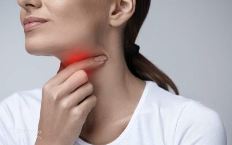
English text: Asymmetrical tonsils, or having one tonsil that looks markedly different from the other, is a classic symptom of tonsil cancer. Another is a persistent sore throat, which does not respond to antibiotics or steroids. Farsi translation: Asymmetric tonsils, that is, when one tonsil is clearly different from the other, is one of the classic signs of tonsil cancer. Another symptom is a persistent sore throat that does not improve with antibiotics or steroid medications.
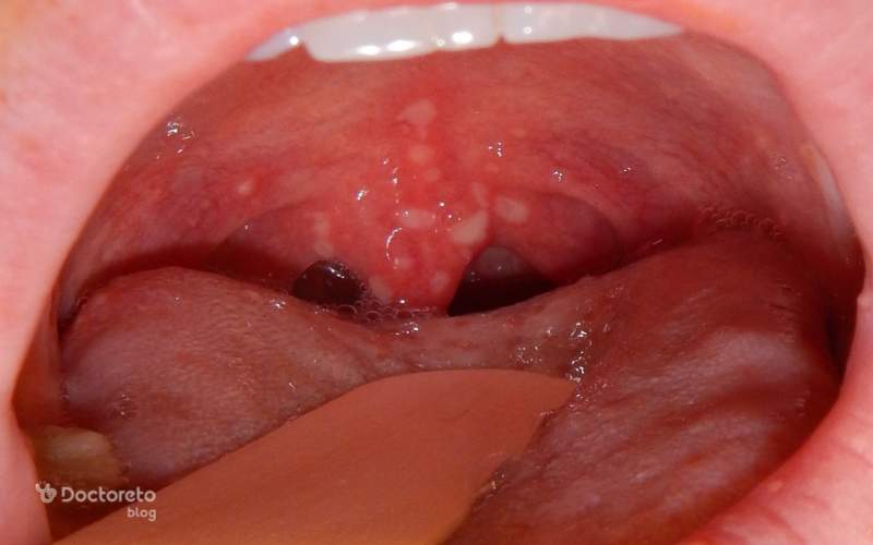
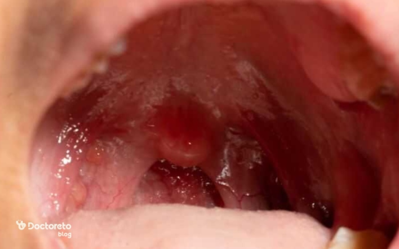
In the early stages, tonsil cancer is usually very subtle and sometimes almost invisible.
In the early stages, tonsil cancer is usually very subtle and sometimes almost imperceptible. At this stage, photos usually show small sores or spots that are mistaken for simple inflammation, which makes people late to notice the disease.
Sometimes the only visible sign is an irritated corner of the tonsil or a small white spot, and no large swelling is seen, but the tonsil tissue becomes rough or bordered, which is detectable to an ENT specialist. The problem with this stage is that the appearance of the lesion is so subtle that one may mistake it for a pest or seasonal inflammation. Therefore, the photos of the initial stage are usually not very clear and only make sense next to other symptoms.
pictures of tonsil cancer in advanced stages
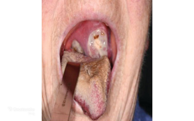
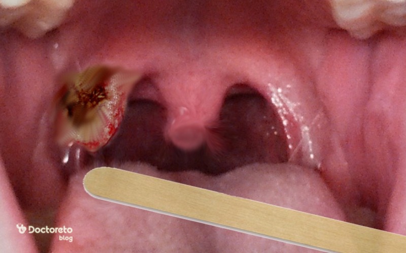
In advanced stages, images usually show obvious changes such as deep ulcers, large masses, unilateral swelling of the throat, and spread to surrounding tissues, which are mistaken for simple inflammation. They don't.
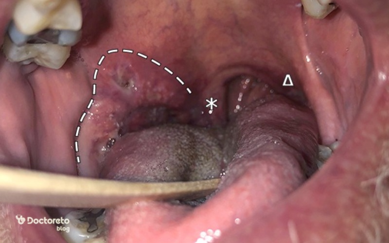
In more advanced stages, the images usually show very clear changes; Deep wounds, large masses, severe unilateral swelling of the throat and sometimes spread to the surrounding tissues are evident in the photographs and are no longer confused with simple inflammation.
In many images, the tonsil lesion is highlighted and may extend to the tongue or throat wall, and in some cases, bleeding or infection-like secretions are seen, which is very worrying, but useful for the doctor's diagnosis. At this stage, the images can lead the doctor to more rapid imaging and a more accurate diagnosis, and the patient usually has symptoms such as ear pain, difficulty swallowing, and bad breath.
English text: The first signs of throat cancer vary depending on where the tumor is. For example, the first signs of oropharyngeal cancer are often a neck lump, ear pain and painful swallowing. A common first sign of laryngeal cancer is hoarseness that doesn't get better. Farsi translation: The initial symptoms of throat cancer differ depending on where the tumor is. For example, in mouth-throat cancer, usually the first symptoms are a lump in the neck, ear pain, and painful swallowing. But in laryngeal cancer, the most common early symptom is hoarseness that does not improve.
photo of the difference between tonsil stones and tonsil cancer
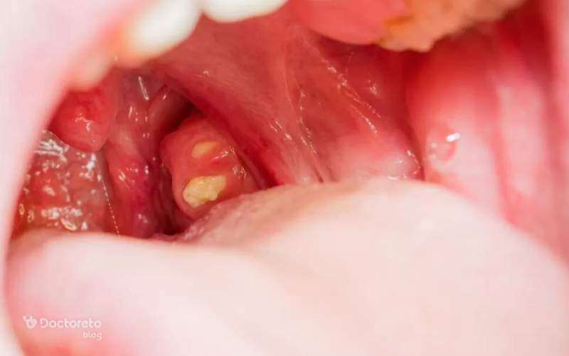
Tonsil stones are usually seen in the form of small white or yellow seeds and are placed on the surface of the tonsil or inside its small cavities.>
Tonsil stones look completely different from tonsil cancer and many comparison photos tonsil stones and cancer show this difference well. Tonsil stones are usually seen in the form of small white or yellow grains and are placed on the surface of the tonsil or inside its small cavities. On images, a tonsil stone is lighter, rounder, and movable, while tonsil cancer appears as a firm ulcer or mass with irregular margins.
Many people are concerned when they see a small white dot, but photos show that these are often tonsil stones, not cancer. These comparison images are reassuring for those who are worried about their symptoms and help them better understand the difference between stones and cancer.
Tonsil stones
Tonsil cancer
It is seen in the form of small white or yellow grains.
It is seen as a sore or fixed mass.
On the surface of the tonsil or inside its small cavities.
It is seen with irregular borders.
It has a completely different appearance from tonsil cancer.
It has a completely different appearance from tonsil stones.
It is brighter, rounder and movable on images.
On images it is a fixed mass with irregular borders.
White and yellow spots are often tonsil stones, not cancer
Many people are worried when they see it.
Comparative photos show this difference well.
Comparative photos help to understand the difference better.
In this table, photos related to tonsil stones and tonsil cancer are reviewed.
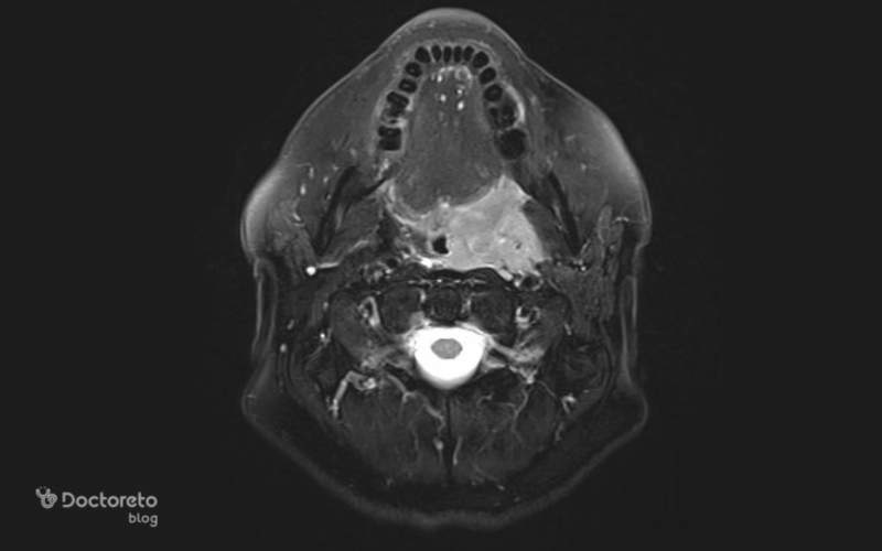
Imaging of tonsil cancer
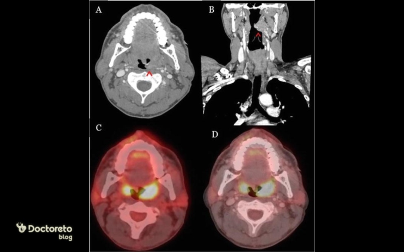
For tonsil cancer imaging, MRI with contrast injection is usually used to accurately determine the extent of soft tissue involvement and CT scan (CT scan) to examine bone involvement and neck lymph nodes.>
The final diagnosis of tonsil cancer is not done only by the appearance of the photo; Rather, medical imaging plays the main role. CT scan, MRI and PET scan are the most common methods. These images help the doctor to check the extent of tissue involvement and possible spread to the lymph nodes or surrounding tissues.
Compared to simple oral photos, medical imaging shows a much more detailed view of the depth and extent of the disease. For example, PET scan is useful to detect the spread of a tumor, but to determine the malignancy of the cells, a biopsy must be performed. Due to this high accuracy, doctors are never satisfied only with the external photo. Imaging is also necessary to determine the stage of cancer and can completely change the course of treatment.
Conclusion
A tonsil cancer photo can be a good starting point for awareness, but should never take the place of a professional diagnosis. The appearance of tonsil cancer can be completely different in different stages and can even be confused with inflammation or tonsil stones. Although seeing the pictures can help calm the mind or even give an early warning, the most important thing is to see an ENT specialist, get a thorough examination and imaging. Any suspicious symptoms that persist for more than two weeks should be taken seriously. In the end, the best way to diagnose and treat is to combine clinical symptoms, careful examination and professional imaging, not relying only on the photos we see on the Internet.
Your doctor takes care of your health!
Frequently Asked Questions
Usually, it is not possible to definitely diagnose tonsil cancer just from a photo. Photographs can show abnormal signs such as a sore or mass, but accurate diagnosis requires a doctor's examination, sampling (biopsy) and pathology tests to determine the type and malignancy of the cells.
No, the stage of tonsil cancer cannot be determined just from the photo. Photographs may show the size or location of the lump, but staging the cancer requires a comprehensive examination, including a physical exam, biopsy, more complete imaging tests (such as CT or MRI), and sometimes lymph node examination.
No, this issue can only be determined by a pathology test.