
Fetal ultrasound in Tehran
Table of Contents
Fetal ultrasound in Tehran
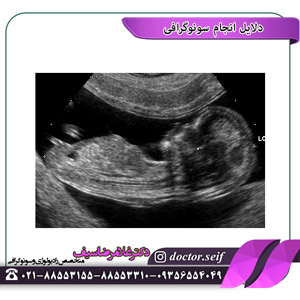
Fetal ultrasound is an imaging technique that uses sound waves to produce images of a fetus in the womb. Fetal ultrasound images can help your gynecologist assess your baby's growth and development and monitor your pregnancy. In some cases, fetal ultrasound is used to evaluate potential problems or help confirm a diagnosis.The first fetal ultrasound is usually done in the first trimester to confirm the pregnancy and estimate how long you are pregnant. If your pregnancy remains uncomplicated, a follow-up ultrasound is usually offered in the second trimester, when anatomical details are visible. If a problem is suspected, a follow-up ultrasound or additional imaging tests may be recommended.
 Fetal ultrasound
There are two main types of fetal ultrasound examination:
sonography Pregnancy
Your doctor may use fetal ultrasound to:
Confirm the pregnancy and its location. Some embryos develop outside the uterus, in the fallopian tubes. A fetal ultrasound can help your doctor diagnose a pregnancy outside the womb (ectopic pregnancy ultrasound).
Determine the gestational age of the child. Knowing your baby's age can help determine your due date and track various milestones throughout your pregnancy.
Confirm the number of babies. If your doctor suspects a multiple pregnancy, an ultrasound may be done to confirm the number of babies.
Assess your child's development. Your doctor can use an ultrasound to determine if your baby is growing at a normal rate. Ultrasound can be used to monitor the child's movement, breathing and heartbeat.
Study the level of placenta and amniotic fluid. The placenta provides nutrients and oxygen-rich blood to your baby. Too much or too little amniotic fluid—the fluid that surrounds the baby in the womb during pregnancy—or placental complications require special attention. Ultrasound can help evaluate the placenta and amniotic fluid around the baby.
Identify birth defects. Ultrasound can help the doctor screen for some birth defects.
Check the side effects. If you are bleeding and have other complications, an ultrasound may help your doctor determine the cause.
Do other prenatal tests. Your doctor may use ultrasound to guide needle placement during prenatal tests, such as amniocentesis or chorionic villus sampling.
Determine the position of the fetus before delivery. Most babies are headless by the end of the third trimester. This does not always happen. Ultrasound imaging can confirm the presentation of the baby so your doctor can discuss delivery options.
Fetal ultrasound should be performed only for valid medical reasons. Fetal ultrasound is not recommended only to determine the gender of the baby. In the same way, ultrasound of the fetus is not recommended only for the purpose of producing videos or photos.
If your doctor does not offer a fetal ultrasound but you want to make sure that an ultrasound can be provided, share your wishes with your provider so that you can work together to determine what is best for you and your baby.
Risks
Diagnostic ultrasound has been used during pregnancy for years and is usually considered safe if used properly. The lowest amount of ultrasound energy that provides an accurate assessment should be used.
Fetal ultrasound also has limitations. Fetal ultrasound may not detect all birth defects – or may falsely indicate a birth defect when it is not present.
For more information on pregnancy ultrasounds, you can contact the radiology center and Dr. Seif's ultrasound or visit Dr. Seif's Instagram. Contact numbers of Dr. Saif's ultrasound center: 88553310
How do you prepare
Depending on the type of ultrasound, you may be asked to drink some fluids or refrain from urinating before the fetal ultrasound. Ask your doctor for instructions when scheduling your ultrasound.
Also note that fetal ultrasound can be performed through the vagina (transvaginal) or upper abdomen (transabdominal), depending on the reason for performing this operation or the stage of pregnancy. If you are having a transabdominal ultrasound, consider wearing loose clothing so that you can easily expose your abdomen.
During the operation
در طی سونوگرافی جنین ترانس شکمی پزشک یا تکنسین شما ژل مخصوصی را روی شکم شما اعمال می کند. This increases the conduction of sound waves and eliminates the air between your skin and transducer.
Your doctor or technician will move the transducer back and forth across your abdomen. The sound waves reflected by your bones and other tissues are converted into images on the monitor.
A doctor or technician will measure your child's anatomy. He may print or save certain images to document important structures. You will most likely be given copies of some of the images.
Depending on the baby's position and stage of development, you may be able to make a face, hands and fingers, or arms and legs. Don't worry if you can't "see" your baby. Ultrasound images are difficult for a trained observer to detect. Ask your doctor or technician to explain what's on the screen.
The method of other types of fetal ultrasound examinations is similar. If you are having an extravaginal ultrasound, you will be asked to change into a patient gown or wedding gown from the waist down. You can sit on the exam table and cross your legs. The transducer is covered in a plastic sheath like a condom and lubricated with gel. Your doctor or technician will insert the catheter into your vagina.
Results
Usually, fetal ultrasound ensures that the baby is growing and developing normally. If your doctor wants more details about your baby's health, he may recommend additional tests.
Other Articles

 Reasons for ultrasound
  sonography Necessary in pregnancy
Answer Cancel %s
study time 6 minutes


