Listen to the summary of this article in this podcast:
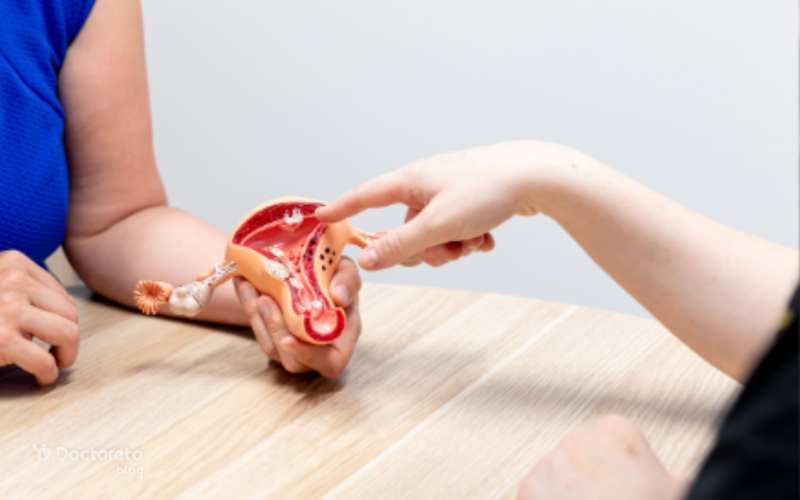
Endometriosis is a female disease in which cells similar to the inner tissue of the uterus grow in areas outside the uterus. Although endometriosis is sometimes seen on ultrasound, diagnosis of endometriosis on ultrasound is not always. If the doctor thinks that the patient has endometriosis, laparoscopic surgery is the only way to definitively diagnose it. Ultrasound for endometriosis helps the doctor to identify the tissue and cysts of endometriosis symptoms. If you have been prescribed ultrasound and you are looking for its interpretation, it is better to consult a gynecologist. You can make an in-person appointment or online consultation with doctors from your doctor's system. If you intend to make an appointment with the best Tehran Laboratory, you can use Doctorto's website.
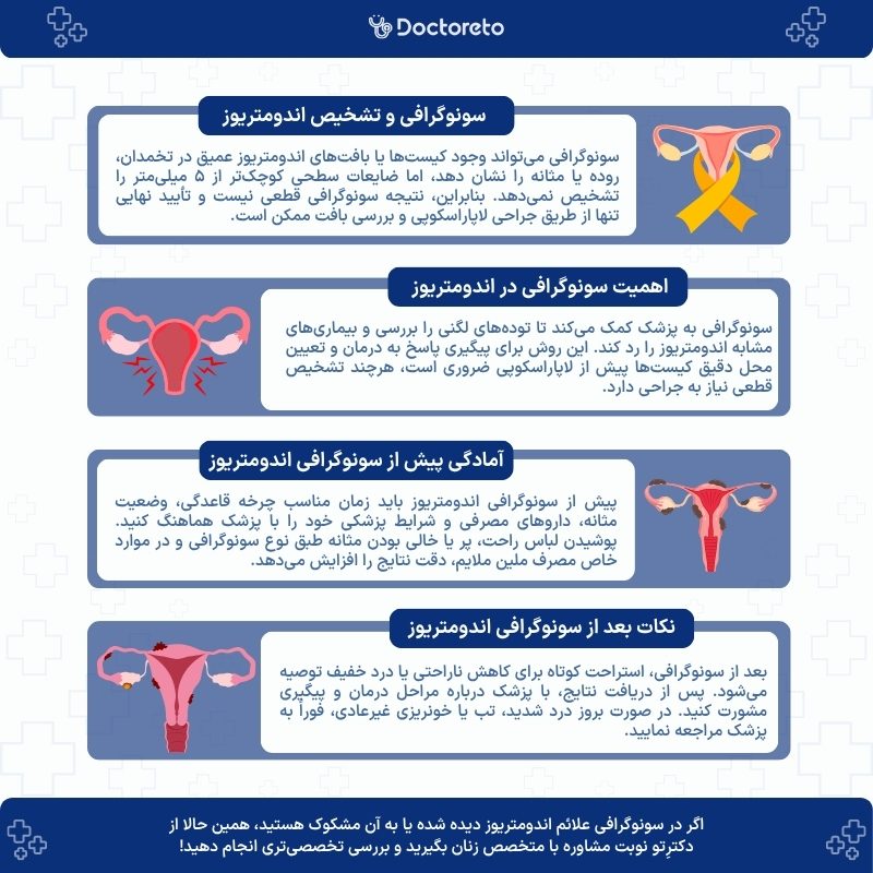
Diagnosis of endometriosis in ultrasound
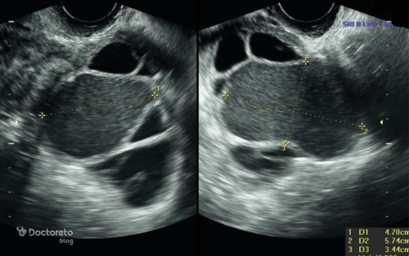
Ultrasound shows large lumps of tissue or sores that are signs of endometriosis. Ultrasounds are mostly used to identify endometriosis in the ovaries; But it cannot show the small pieces of tissue that are the most common type of endometriosis. In fact, the diagnosis of endometriosis in ultrasound is not very certain, and this test can show the endometrial tissue that has turned into cysts (endometriums) and is mostly found in the ovaries.
Endometrial tissue that has penetrated deep into the intestine or bladder can also be detected through ultrasound. But tissues Endometriosis A surface smaller than 5 mm is not seen in ultrasound, which is the most common type of endometriosis. The important and complicated thing about the diagnosis of endometriosis in ultrasound is that even if the ultrasound shows a tissue that does not belong to it, it cannot be said definitively that it is endometriosis. The only way to be sure is to remove tissue and test it, which is done with laparoscopic uterine operation.
Ultrasound is suggested to be performed at the end of the menstrual period and when the inner tissue of the uterus is the thinnest (day 4 to 9 of the menstrual cycle)
necessity of doing endometriosis ultrasound
Ultrasound is only one medical tool to determine if you have endometriosis. This method helps the doctor rule out diseases with symptoms similar to endometriosis, or can more closely examine the mass observed in the pelvic exam. In fact, the necessity of doing an endometriosis ultrasound is what brings the doctor closer to the right treatment in the later stages of treatment.
You don't need an official diagnosis to receive common endometriosis treatments, such as birth control pills and progesterone, which help reduce symptoms, and you can monitor the cyst's response to treatment with an endometriosis ultrasound. In addition, by performing an endometriosis ultrasound, the doctor finds out where the cyst and endometriosis are located so that he can perform the laparoscopy more easily.
Infographic of endometriosis diagnosis in ultrasound
Types of ultrasound to diagnose endometriosis
Diagnosis of endometriosis is usually done using different imaging methods such as ultrasound. Ultrasound can help identify endometriosis cysts (endometrioma) and check the status of ovaries and other pelvic organs. Different types of ultrasound include transabdominal ultrasound (through the abdomen) and transvaginal ultrasound (through the vagina). Transvaginal ultrasound is more accurate and is usually used to more accurately evaluate endometriosis because it allows the doctor to better see tissues and changes in deeper areas of the pelvis. The types of ultrasound that are used to diagnose endometriosis are:
Type of ultrasound
Description
Transvaginal ultrasound (TVS)
This type of ultrasound is performed by inserting an ultrasound probe into the vagina and provides a clearer picture of the internal organs of women, including the ovaries and uterus, and cysts and It detects abnormal tissues.
Abdominal ultrasound
In this procedure, the ultrasound probe is placed on the abdomen and detects large masses and cysts, but it does not provide a clearer picture than transvaginal ultrasound.
Rectal ultrasound or transrectal ultrasound
One of the Specialized methods for diagnosing endometriosis are ultrasounds, especially in cases where endometriosis has penetrated the intestines. In this procedure, the ultrasound probe is inserted through the rectum to produce detailed images of the pelvic area and intestines.
Endometriosis Mapping Ultrasound
It is an advanced method to identify and accurately map areas affected by endometriosis, especially in cases where there is deep infiltrating endometriosis.
Types Ultrasound to diagnose endometriosis
English text: Although laparoscopy is the gold-standard test to establish a diagnosis of deep endometriosis, transvaginal ultrasound (TVUS) can contribute to its detection, because it is an accessible, noninvasive examination that allows preoperative planning in cases requiring surgical treatment. Text translation: Although laparoscopy is the best method for diagnosing deep endometriosis, ultrasound Transvaginal (TVUS) helps to identify it. This method is a type of non-invasive and accessible examination that allows the doctor to make the necessary plans before surgery in cases where surgical treatment is needed.
1. Diagnosis of endometriosis with vaginal ultrasound
Diagnosis of endometriosis with vaginal ultrasound is one of the effective methods of diagnosing endometriosis in ultrasound, which gives the doctor accurate images of the internal organs, especially the ovaries and uterus. One of the most important benefits of vaginal ultrasound is that it provides clearer images of soft tissues that help the doctor identify cysts and abnormal tissues. Also, this method has the ability to identify endometrial cysts (endometriomas) and other lesions related to endometriosis.
However, diagnosing endometriosis with vaginal ultrasound also has limitations; Because this method cannot identify all types of endometriosis, especially superficial endometriosis or very small lesions. In general, vaginal ultrasound is a valuable tool in the diagnosis and management of endometriosis, which helps identify this disease more quickly and effectively.
In abdominal ultrasound, the ultrasound probe is placed on the abdomen and detects large masses and cysts.
2. Diagnosis of endometriosis with abdominal ultrasound
Diagnosis of endometriosis with abdominal ultrasound is one of the common ways to identify this disease. In this method, the ultrasound probe is placed on the abdomen to take images of the internal organs to identify large cysts and abnormal masses. Although this method is effective in identifying endometriosis in the ovaries, due to the type of imaging and the distance from the organs, its results may not be as accurate as transvaginal ultrasound.
This method can be used as part of the initial evaluation to diagnose endometriosis and check the general condition of the pelvis. Abdominal ultrasound results help the doctor in planning the next steps of treatment, but for a definite diagnosis and identification of different types of endometriosis, it may be necessary to perform other methods, such as transvaginal ultrasound or laparoscopic surgery.
3. Rectal ultrasound for endometriosis (transrectal ultrasound for endometriosis)
Rectal ultrasound for endometriosis allows doctors to more closely examine deeper tissues and lesions in the areas around the intestines. Transrectal ultrasound is especially effective in identifying deep infiltrating endometriosis in the intestinal wall. The advantage of this method is in the detailed information it provides about the state of endometriosis in sensitive areas close to the intestines and helps the doctor in planning treatment and deciding on the type of surgery needed. However, rectal ultrasound for endometriosis as a specialized method may not be necessary in all cases and is usually recommended in certain situations when the doctor suspects the presence of endometriosis in the intestines.
4. Endometriosis mapping ultrasound
Endometriosis mapping ultrasound maps the pelvic area to determine the position, size and depth of endometriosis. The importance of these key details cannot be overlooked. This information helps the doctor to have a precise understanding of the condition of endometriosis and to plan a better treatment. Accurate identification of endometriosis areas can reduce surgical risks and improve treatment results. Endometriosis mapping ultrasound provides a closer view of the organs affected by endometriosis such as the ovaries, bladder and intestines.
The best ultrasound time to diagnose endometriosis
Pelvic ultrasound can be performed at any stage of a woman's menstrual cycle. To achieve the best imaging results, it is suggested for women who have not finished their menstrual period yet that the ultrasound be performed at the end of the menstrual period and when the inner tissue of the uterus is at its thinnest (day 4 to 9 of the menstrual cycle). The timing of the ultrasound is not critical, but if the problem you are experiencing is heavy bleeding, the best time for an ultrasound to diagnose endometriosis is right after your period ends. When going to the ultrasound, the patient is asked to fill out a form related to her femininity history.
Get to know how to diagnose endometriosis in ultrasound.
Preparation before endometriosis ultrasound
Preparation before endometriosis ultrasound improves the quality of the images and the accuracy of the results. You can prepare for this test by observing the following points:
Before the ultrasound, be sure to consult your doctor about any concerns or questions you have.
When visiting, fill out the forms related to your medical history and femininity. This information helps the doctor to better understand what problems you have.
If you are still menstruating, it is better to do the ultrasound on days 4 to 9 of the menstrual cycle so that the inner tissue of the uterus is thinner.
For transvaginal ultrasound, the bladder must be empty. In some ultrasounds, the doctor may recommend that you drink some liquids to fill your bladder before the ultrasound.
Wear comfortable and loose clothes to be more comfortable during the ultrasound.
Be sure to tell the ultrasound technician if you are taking certain medications or have any other medical conditions.
Some doctors prefer the patient to have a mild bowel preparation before the ultrasound; Therefore, he prescribed a mild laxative that should be taken the night before the ultrasound. This is usually done when you have a history of severe endometriosis or feel significant pain in your bowels during your period.
English text: You will need to be well hydrated prior to the procedure and may drink water throughout the day, but you will not need a full bladder. Please do not eat for 2 hours prior to your appointment time. Translation of the text: Before the ultrasound, it is necessary that the body is well hydrated. The patient can drink water during the day, but it is not necessary to fill the bladder completely. Do not eat for 2 hours before the ultrasound.
An endometriosis ultrasound can show large clusters of tissue or lesions that may be signs of endometriosis.
Important points after endometriosis ultrasound
After performing an endometriosis ultrasound, following some important points can improve the patient's condition and facilitate the follow-up process. Rest is very important after ultrasound. If you feel discomfort or mild pain, it is better to rest for a while to get better. After receiving the results, consult with your doctor about the next steps of treatment or further investigations to better plan your endometriosis management.
It is also very important to pay attention to the symptoms after ultrasound. If you have unusual symptoms after the ultrasound, such as heavy bleeding, severe pain, or fever, see your doctor immediately. These symptoms are signs of side effects that need to be investigated. If the doctor has given a special order after the ultrasound, be sure to follow it.
Probability of error in endometriosis ultrasound
Standard ultrasound cannot confirm whether a person has endometriosis or not, but it can identify cysts associated with this disease, which are called endometriomas. An endometriosis ultrasound can show large clusters of tissue or lesions that may be symptoms of endometriosis. This method is very effective in identifying endometriosis in ovaries. However, ultrasounds cannot detect the small pieces of tissue that are the most common type of endometriosis. Currently, laparoscopic surgery is the only definitive way to confirm the diagnosis of endometriosis.
endometriosis ultrasound price
The price of endometriosis ultrasound depends on various factors, which include:
Place of ultrasound: Endometriosis ultrasound costs are higher in hospitals, clinics or private centers.
Type of ultrasound: Transvaginal ultrasound usually has a different cost than abdominal ultrasound.
Health insurance: if you are covered by insurance, a part of the ultrasound costs will be covered.
Geographical location: prices vary depending on the geographical area; In big cities, the cost of ultrasound for endometriosis is higher.
For more information about the price of endometriosis ultrasound, contact the center that is going to perform this test.
Conclusion and guide to see a doctor
Diagnosis of endometriosis in ultrasound is an effective tool in the management of this disease. Transvaginal ultrasound can identify cysts and abnormal tissues of endometriosis, but laparoscopic surgery is required for definitive confirmation. If you have symptoms such as severe pelvic pain or abnormal bleeding after the ultrasound, be sure to see a gynecologist. Fortunately, you can get help from your doctor's online consultation service for this purpose.
Your doctor takes care of your health!
Frequently Asked Questions
The best way to diagnose endometriosis, and the only definitive way to confirm the presence of endometriosis and check its depth and extent, is to perform laparoscopic surgery.
Women who have severe pelvic pain, abnormal bleeding, or pain during menstruation are experiencing, are trying to get pregnant and have a history of fertility problems, have a family history of endometriosis and ovarian cysts, or endometrial cysts (endometrium) detected by ultrasound should be examined and further tests for endometriosis.
Yes, it is possible to diagnose endometriosis without ultrasound, but it is more complicated. The doctor finds out the possibility of endometriosis by examining the medical history, clinical symptoms and physical examination. In some cases, other tests such as an MRI or biopsy may be needed. But for definitive confirmation and accurate evaluation, ultrasound or laparoscopic surgery is usually necessary.
I have started my activity in the field of content production and management since 1995 and I am always eager to face new job opportunities. Getting to know a different world in different fields such as medicine and health also encourages me to follow a healthy lifestyle. That's why I started working with your doctor.