
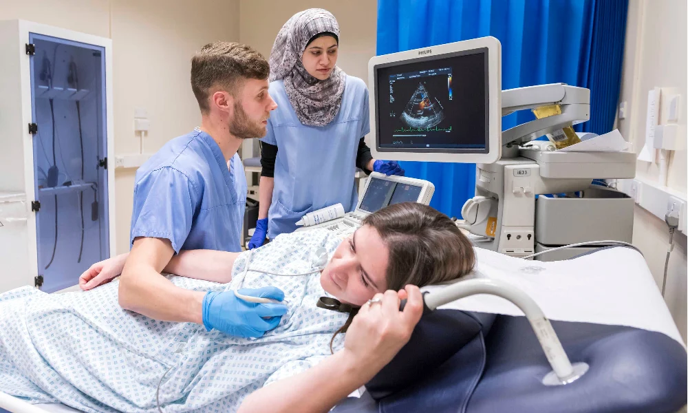
Women's heart echo The importance of women's echo and its differences with men's heart echo
What is the difference between women's heart echo and men's echo?
Nowadays, women may need an echocardiogram for several reasons, including congenital heart diseases, pregnancy complications, and evaluation before and after pregnancy-related surgeries. Echocardiography helps medical professionals to take a comprehensive look at the structure, function and blood flow in the heart. In this article, we will introduce you to what everyone is asking about women's heart echo.
Easy accessShare content
Instagram Facebook X-twitter TelegramWhat is women's heart echo? How is it different from Echo Men?
Actually, heart echo or echocardiography is a safe imaging method in which moving images of the heart are created using high-frequency sound waves. Although echocardiography of both men and women is used for the same purpose and to diagnose and evaluate heart diseases, due to the anatomical differences between men and women, there are small differences in how it is done.
For example, in some cases, due to the difference in body shape, the echo probe may have a different positioning angle when performing it for women and men, or due to the presence of breasts in women's organs, it is possible to use certain positions in women's heart echo to receive clearer images of the heart.
Familiarity with the types of women's heart echo
Generally, echocardiography is done in two ways:
Transthoracic echocardiography
This echo is known as the most common type of echo that is done through the chest. This method involves first applying a lubricating gel on the chest and then moving a probe or transducer on the chest. This transducer sends sound waves to the heart and the reflection of these waves is seen as an image on the screen.

Transesophageal echocardiography
This method is a bit more difficult than the previous method. During this procedure, instead of contacting the chest, the probe enters the body through the mouth and esophagus and approaches the heart. This method is recommended if there is a need to get more detailed images of the heart, especially the back part of the heart.
Obviously, consulting a specialist doctor to choose the right type of echocardiography to diagnose your disease will help you get a better result and access faster treatments.
Advantages of women's heart echo
After the ECG test, echocardiography is a common method prescribed by the doctor to check and diagnose heart diseases. Among the benefits of echocardiography for women, the following can be mentioned:
- Non-invasiveness: Echocardiography is a painless method and does not require the injection of contrast material.
- Safety: The sound waves used in echocardiography do not pose any danger to the body.
- High accuracy: Echocardiography provides very detailed images of the heart.
- High speed: The duration of echocardiography is usually short and does not take much time.
- Reasonable cost: This method is less expensive than other imaging methods.
How is a women's heart echo performed?
Since echo is a simple, short and safe method, it also includes simple steps. respectively:
- Preparation: First, the patient takes off his upper body clothes and lies on his back on the bed.
- Gel application: A lubricating gel is applied on the chest to make the transmission of sound waves through the skin smoother and better.
- Probe placement: The doctor or ultrasound technician moves the probe or transducer over the chest.
- Imaging: Sound waves are sent through to the heart and their reflection is seen in the form of images of the heart on the monitor.
- Interpretation of the images: After viewing the images, the doctor examines them and obtains useful information about the condition of the patient's heart.
What happens during echocardiography?
You may be asked to take deep breaths during the echocardiogram. This helps to get better pictures of the heart. You may need to turn to the left or right so that the doctor can see and examine your heart from different angles, or you may need to hold your breath for a few seconds. This allows for clearer images of the heart valves.
What are the preparation steps before echocardiography for women?
As it has been repeatedly said, women's echocardiography is a simple, safe and painless procedure and usually does not require any special preparation, but in some cases, recommendations by the doctor may be considered for you. Things like clothing that can be easily opened from the waist up and avoiding jewelry and other metal objects are among these recommendations.
Fasting before women's heart echo
Generally, there is no need to fast to perform a regular echocardiogram, but if the echocardiogram is prescribed through the esophagus, you should fast for 8 hours before doing it and avoid eating and drinking. The doctor's order is decisive in the case of stopping or taking medications before both types of women's echocardiography.
When is it necessary to perform echocardiography?
There are several cases that compel the doctor to prescribe echocardiography for women. Among them, the following situations can be mentioned:
- Before pregnancy: Women's heart echo is necessary to check the heart health of women who are planning to become pregnant and have a history of heart disease or high risk factors such as blood pressure, diabetes, and obesity.
- During pregnancy: Doing an echocardiogram to monitor the health of the heart of the mother and the fetus is prescribed by the doctor in women who face symptoms such as shortness of breath, fatigue, or leg swelling during their pregnancy, or who suffer from heart diseases.
- After delivery: Some women may experience heart changes or possible complications due to the delivery process. It is necessary to do echo for these women to check these things.
- If there are symptoms: In case of symptoms such as chest pain, shortness of breath, palpitations, dizziness or swelling of the legs, the doctor may prescribe an echocardiogram.
- As part of screening: To screen for heart disease in women who have risk factors.

How long does a women's heart echo take?
Generally, echocardiography is a fast method, but in some cases, the time required to perform an echocardiogram varies according to its type and the condition of the patient. Usually, heart echo from the chest, as a common type of echo, requires about 15 to 30 minutes, while esophageal echo takes a little longer and about 30 to 45 minutes due to the preparation and placement of the probe in the esophagus.
In some cases, the doctor, feeling the need for a more detailed examination, takes more images from different angles of the heart, which also leads to an increase in echo time. Another point is the cooperation or non-cooperation of the patient during the echo, which affects the duration of the echo. Finally, it is obvious that the skill of the specialist doctor who performs this test has a significant impact on the quality and time of the echo. Performing echocardiography by an experienced doctor with a brilliant record gives you confidence and peace of mind in all stages of diagnosis and treatment.
What is determined in women's heart echo?
Echocardiography is a method that uses images created by sound waves to evaluate the structure of the heart and the health of blood flow. Performing an echocardiogram provides the following set of information to a specialist doctor.
- Size and shape of the heart: Is the heart enlarged or is its shape abnormal?
- Thickness of the heart walls: Are the heart walls thickened or thinned?
- Function of heart valves: Do the valves open and close properly?
- Blood flow in the heart: Is the blood flow in the heart normal or is there a blockage?
- Size of the heart chambers: Are the heart chambers enlarged or reduced?
- Presence of a blood clot: Is there a blood clot in the heart?
- Cardiac tumors: Is there a tumor in the heart?
- Congenital defects of the heart: Is the heart associated with congenital defects?
- Injury to the heart muscle: Is the heart muscle damaged?
- Blood pressure in the blood vessels around the heart: Is the blood pressure in the blood vessels around the heart normal?
What does the echo of the heart show?
After performing echocardiography, the doctor, by observing and interpreting the resulting images, obtains important information that helps to accurately diagnose the condition of the patient's heart and make the best decision for treatment. Identifying things like:
- valvular heart diseases: such as narrowing or leaking valves
- Heart muscle diseases (Cardiomyopathy): Abnormal thickening, thinning or stiffening of the heart muscle
- Congenital heart defects: Heart problems that are present from birth
- Heart rhythm problems (arrhythmia): irregular heartbeat
- Heart failure: inability of the heart to pump enough blood to the body
- Blood clots in the heart: which can lead to stroke
- Cardiac tumors: Abnormal growth of cells in the heart
In addition to diagnosing the above diseases, women's heart echo helps the doctor to:
- Determine the severity of the disease.
- Evaluate the effectiveness of the treatment.
- Follow the progress of the disease.

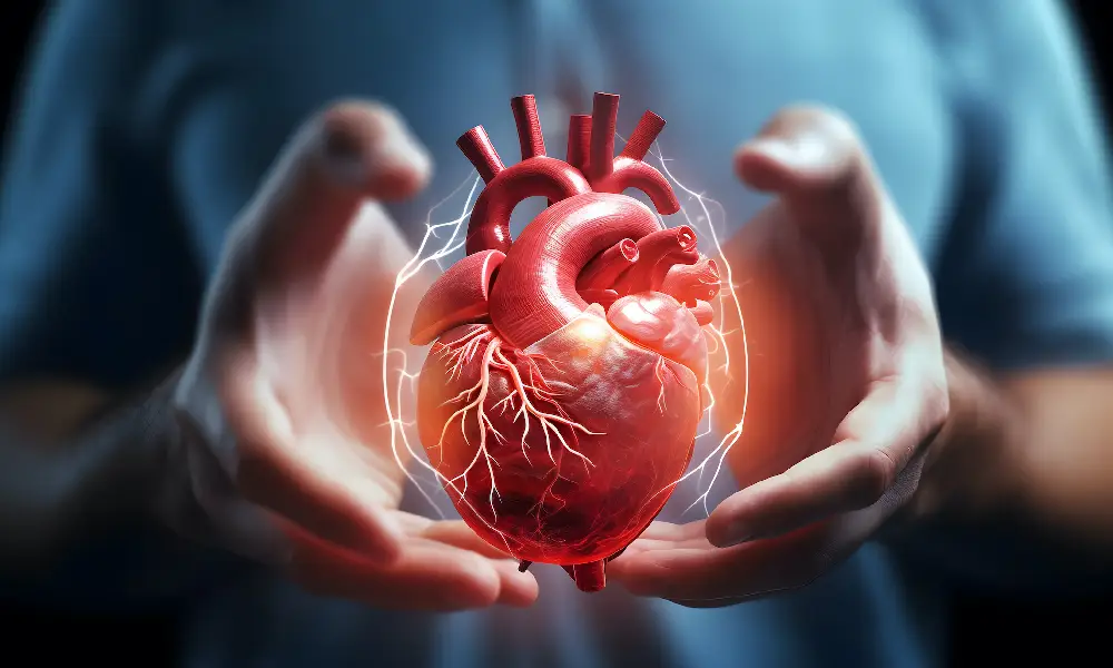
Why is echocardiography important for women?
Heart echo is a very important method to check heart health. Echocardiography of women is important in many ways. Like:
hormonal changes
Women experience a lot of hormonal fluctuations during menstruation, pregnancy and after menopause, and these fluctuations can increase the risk of heart diseases by affecting the functioning of the heart and blood vessels.
Pregnancy
The beginning of pregnancy and during it causes many important changes in women's body. Among these changes, we can mention an increase in blood volume and changes in blood pressure, all of which increase the need for more investigation and monitoring during pregnancy due to their impact on the activity of the heart and blood vessels.
Congenital heart diseases
Some women are suffering from heart diseases from birth, and these diseases may not cause any problems during their life, but during pregnancy or menopause, there is a possibility of their aggravation.
risk factors
In women, factors such as high blood pressure, diabetes, high cholesterol and obesity that increase the risk of heart diseases are common, and these factors also make the need for cardiac examinations necessary. Edit Clone Comments read more Why care about leg veins? Solutions to prevent vascular problems
02
April
Why care about leg veins? Solutions to prevent vascular problems
02
April
 10 effective ways to maintain heart health and prevent cardiovascular diseases
02
April
10 effective ways to maintain heart health and prevent cardiovascular diseases
02
April
 introduction of several home treatment methods for varicose veins
07
D
introduction of several home treatment methods for varicose veins
07
D
 Preparation before taking an ECG; Should we be fasting to take an ECG?
07
D
Preparation before taking an ECG; Should we be fasting to take an ECG?
07
D
 The least dangerous methods of treating coronary artery occlusion
07
D
The least dangerous methods of treating coronary artery occlusion
07
D
 3-day apple weight loss diet
07
D
Your comments
0
0
votes
Article Rating
Subscribe to
Login
0 Comments
the oldest
the most recent
The most votes
Feedback (Feedback) inline
View all comments
3-day apple weight loss diet
07
D
Your comments
0
0
votes
Article Rating
Subscribe to
Login
0 Comments
the oldest
the most recent
The most votes
Feedback (Feedback) inline
View all comments


