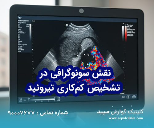
Ultrasound for diagnosing hypothyroidism
The thyroid, this butterfly-shaped gland at the front of the neck, despite its small size, is the master of the body's metabolism. When it comes to hypothyroidism, many think that only a blood test (TSH) is enough to make a diagnosis. But in the new medical methodologies of 2026, an endocrinology and metabolism doctor relies on a more accurate tool called thyroid ultrasound to understand the root cause of the disease and predict the treatment process. Alone is not enough?
Biochemical tests only show the functional state of the gland, that is, they tell us how much hormone the thyroid is currently producing. But these tests are not able to reveal structural changes, hidden inflammations or presence of suspicious nodes. Ultrasound, as the main supplement, places "anatomy" next to "function". In cases where the patient presents with classic symptoms of hypothyroidism, such as excessive fatigue, weight gain, and dry skin, but the blood test is in the "subclinical" range, ultrasound can show tissue changes caused by immune system invasion (such as Hashimoto's) long before the sharp drop in hormones. gform_wrapper gravity-theme gform-theme--no-framework" data-form-theme="gravity-theme" data-form-index="0" id="gform_wrapper_8">
Specific signs of hypothyroidism in ultrasound
In a specialized center, a radiologist looks for certain patterns when examining a patient with hypothyroidism:
- Decreased echogenicity (Hypoechogenicity): Normal thyroid tissue can be seen in clear ultrasound. In inflammatory diseases that lead to hypofunction, the tissue appears darker than normal, indicating replacement of healthy cells with lymphocytes and fibrotic tissue.
- Irregular and lobulated tissue: The edges of the thyroid gland are normally smooth. In chronic hypofunction, these edges become wavy. Vascular changes: Using color doppler technology, the doctor examines the amount of blood supply to the gland. In the early stages of inflammation, the blood supply may be greatly increased (Thyroid Inferno), which indicates an overactive immune system against the gland.
ulsonography findings in types of hypothyroidism (2026 version)
| Type of disorder | Gland size in ultrasound | Echogenicity status (tissue) | Blood supply status (Doppler) | Recommendation of endocrinology and metabolism doctor |
|---|
| Hashimoto's thyroiditis (stages) primary) | Larger than normal (goiter) | Heterogeneous (incoherent) and dark | Severe increase (Hypervascular) | Initiate drug therapy and antibody monitoring |
| Atrophic hypothyroidism | Very small and contracted | Very dark and Fibrotic | Severe reduction of blood supply | Permanent hormone replacement (levothyroxine) |
| Secondary (central) hypofunction | Normal or small | Homogeneous and normal tissue | Normal | Pituitary gland examination and MRI Cerebral |
| Postpartum thyroiditis | Large and swollen | Local reduction of echogenicity | Variable | Periodic follow-up (usually transient) |
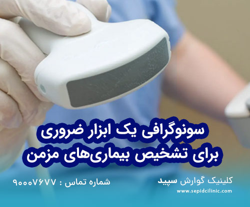
Importance of choosing ultrasound center in Shahrek Gharb Tehran
The accuracy of Diagnosis of thyroid diseases in ultrasound strongly depends on two factors: the technology of the device and the skill of the radiologist. In an area like West Tehran, due to the high density of medical centers, choosing a Elastography allows the doctor to measure the stiffness of the thyroid tissue. Tissues that are fibrosed by chronic underactivity are stiffer. These data help the doctor of endocrinology and metabolism to more accurately determine the prognosis of the disease. Does the gland still have the potential to return to normal function or is the structural damage permanent?
Investigation of thyroid nodules in the bed of hypothyroidism
One of the big challenges in patients with hypothyroidism is the high prevalence of nodules or thyroid nodules. Chronic inflammation in Hashimoto's thyroiditis can give rise to nodules that are difficult to distinguish from malignant tumors on conventional ultrasound.
In advanced centers, use of the TI-RADS classification system (updated version 2026) is mandatory. This system allows the radiologist to estimate the risk of malignancy based on the shape, margin, presence of microcalcification (calcium deposits) and internal composition of the nodule. If you undergo ultrasound at a reputable center in West Tehran, your report will include accurate TI-RADS scoring, which will prevent unnecessary biopsies (FNA). rel="dofollow" class="uc406ce42a20e51b935cc2233dc2826d1">
You may also be interested in this article... Comprehensive guide to diagnosis and treatment of hypothyroidism
biological effects and scientific solutions for disease management
Hypothyroidism is not just a hormonal drop, but a change in the biochemistry of the whole body. From a scientific point of view, when the thyroid gland is under inflammatory stress (appearing on ultrasound with a "holed" or moth-eaten appearance), lifestyle management is as important as medication:
- Stress management: High cortisol directly inhibits the conversion of T4 to T3 (the active form of the hormone).
- Anti-inflammatory nutrition: Selenium and zinc intake (under consideration doctor) can help reduce tissue inflammation seen in ultrasound.
- Avoid excessive fluoride and chlorine: These elements can replace iodine in the thyroid gland and accelerate the tissue destruction process.
Doppler ultrasound, a new port toward treatment More precisely
Many patients ask: "Why is color ultrasound (Doppler) prescribed for simple hypothyroidism?" The answer lies in "blood flow assessment". In lymphocytic thyroiditis, the vessels of the gland become dilated due to the inflammatory response. Observing this pattern assures the endocrinologist and metabolism doctor that the cause of hypofunction is autoimmunity and not iodine deficiency or other issues. This differential diagnosis completely changes the treatment protocol.
Preparations before referring to the ultrasound center
In order for your ultrasound to be performed with the most accuracy in the ultrasound center of Shahrek Gharb Tehran, it is recommended to observe the following:
- Avoid wearing clothes with a ski collar or bulky jewelry.
- There is no need to fast, but it is better to avoid coffee or strong stimulants that increase the heart rate before the test (so that it does not affect the doppler). Comparing "gland volume changes" over time is the most valuable data for your doctor.
Summary and final word of the role of ultrasound in the diagnosis of hypothyroidism
Finally, it should be remembered that the successful management of hypothyroidism in 2026 is more than taking a daily pill, it It is a multi-pronged approach where detailed imaging maps the road map. Ultrasound, as the "eye" of the doctor of endocrinology and metabolism, allows him to be aware of the inflammatory or structural nature of the disease and prevent complications such as suspicious nodes or complete atrophy of the gland. Visiting an ultrasound center in West Tehran, using advanced technologies such as elastography, gives you the assurance that the smallest tissue changes are not hidden from the experts and your treatment path is designed based on the most accurate biological data. Thyroid health is the foundation of your daily energy and vitality, so make close structural monitoring a priority on par with blood tests.
Frequently asked questions (FAQ) about hypothyroidism
1. Does thyroid ultrasound have radiation and is it dangerous?
No, ultrasound uses sound waves and does not contain any x-rays or ionizers. Doing it is completely safe even for pregnant women and children.
2. If the ultrasound is normal but the TSH is high, what should be done?
In this case, the diagnosis is usually "primary hypofunction without structural change". This means that the gland is not damaged yet, but is lazy in producing hormones. The treatment is started based on the hormone level by the endocrinology and metabolism doctor.
3. How often should the ultrasound be repeated?
If nodules are present, usually every 6 to 12 months. For simple hypothyroidism without nodules, annual ultrasound or once every two years is enough to check the atrophy process (analysis of gland loss).
4. Can ultrasound diagnose thyroid cancer definitively?
Ultrasound determines the "likelihood" of malignancy (via the TI-RADS system). Definitive diagnosis is possible only through needle biopsy (FNA) which is performed under ultrasound guidance.
5. Why is ultrasound recommended in the specialized centers of Shahrek Gharb?
Due to access to ultra-advanced devices with high frequency (High Frequency) that are able to detect nodes smaller than 2 mm and subtle tissue changes, it is a priority to refer to reputable centers in this region. Do:
American Thyroid Association (ATA): thyroid.org – the world's leading authority on thyroid treatment guidelines.
RadiologyInfo.org: The complete guide to thyroid ultrasound for patients.
The Lancet Diabetes & Endocrinology: The latest research articles on Hashimoto's epidemiology and new imaging techniques.
For an appointment, just contact us.
Free consultation number :
09305576635
90007677 90007677 Do you agree?","legend":"0\/5 - (0 points)","size":"24","title":"The role of ultrasound in diagnosing hypothyroidism","width":"0","_legend":"{score}\/{best} - ({count} {votes})","font_factor":"1.25"}'>